You are viewing 1 of your 1 free articles
Masterclass: SLAP lesions - Part I
In part one of this two-part article, Chris Mallac explains the essential anatomy and biomechanics of the biceps anchor, how and why injury occurs, and how to diagnose an injury.
Chicago White Sox starting pitcher Dylan Covey (68), 2018. Credit: Kamil Krzaczynski-USA TODAY Sports
The SLAP lesion was recognized as being a definitive clinical entity after the development of shoulder arthroscopic interventions(1). SLAP is an acronym for ‘superior labral tear from anterior to posterior,’ and the term ‘SLAP’ lesion refers to a particular shoulder injury that affects the long head of the biceps anchor to the glenoid labrum.
The understanding of the pathophysiology behind SLAP lesions has progressed greatly since the advent of arthroscopic shoulder surgery. Previously, with open-shoulder surgery, it was difficult to visualize the superior labrum and biceps attachment, resulting in unknown, missed, and poorly understood SLAP lesions.
Although injuries to the superior glenoid labrum are uncommon(2), an injury to the superior labrum and biceps anchor can be a source of deep shoulder pain, and it is frequently seen as an overuse injury in overhead athletes and baseball pitchers. If left untreated, a SLAP lesion can have devastating consequences for the overhead athlete in terms of pain, dysfunction, and loss of power in sports.
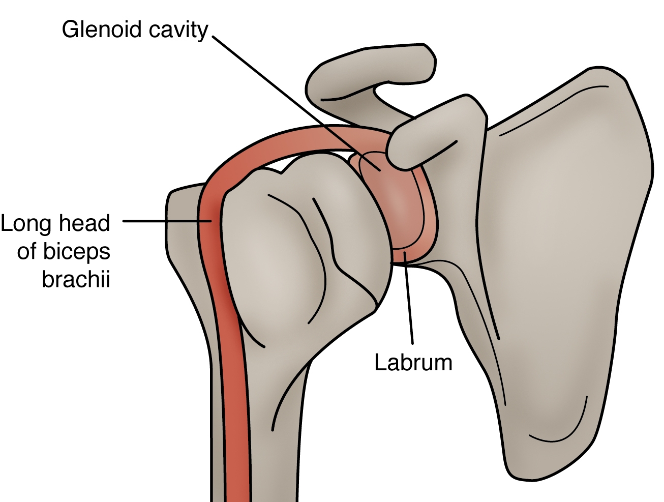
Figure 1: Anatomy and biomechanics of GHJ
Anatomy and biomechanics (see figure 1)
The glenohumeral joint (GHJ) relies on passive and active restraints to create joint stability. The passive restraints include the capsule-ligamentous structures, the glenoid labrum, and negative intra-articular pressure. Dynamic restraints include rotator cuff muscles, periscapular muscles, and the long head of the biceps muscle, which attaches to the superior margin of the labrum and glenoid rim(3).
Of particular interest is the fibrocartilage labrum that surrounds the glenoid rim circumferentially, increases the depth of the glenoid socket, and enhances the passive stability of the GHJ(4). Expanding the glenoid fossa’s depth reduces the shear effect of the humeral head (5). The superior part of the labrum is loose and meniscoid in structure and attached by loose connective fibers to the glenoid rim. Up to 50% of the long head of the bicep tendon originates on the superior labrum, with the remaining 50% attached to the supraglenoid tubercle(4).
It is well understood that the rotator cuff works as a force couple to contract and compress the humeral head into the glenoid fossa during shoulder movements. The long head of the bicep also plays a role in ensuring GHJ stability. The long head of the biceps tendon and superior labrum help stabilize the humeral head (usually in the abducted and externally rotated arm) through a pulley action, which is an important requirement in sports such as baseball pitching. It is reported that complete lesions of this biceps-labrum complex result in a 6mm increase in GHJ anterior translation(6).
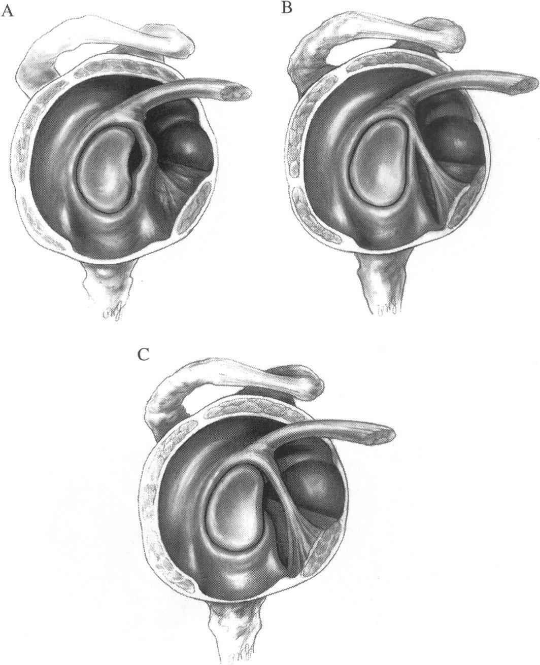
Figure 2: Anatomical variants of the glenoid labrum
Normal anatomic variants of the anterosuperior glenoid labrum and glenohumeral ligaments. A = Sub-labral foramen; B = Cord-like middle glenohumeral ligament; C = Buford complex (cordlike middle glenohumeral ligament without anterior superior labral tissue). From Alessandro et al 2000
Interestingly, the glenoid labrum has some major anatomical variants (see figure 2). The primary ones of interest, and which should not be confused with a genuine SLAP lesion, are(5):
- Sub-labral foramen is a groove between the normal anterosuperior labrum and the anterior cartilaginous border of the glenoid rim. They are usually seen in the 3-o’clock position.
- A small sub-labral recess represents a gap located inferior to the biceps anchor and the anterosuperior portion of the labrum. It is usually seen in a 12-o’clock position of the glenoid in arthroscopic surgery.
- Cordlike middle glenohumeral ligament.
- Buford complex, characterized by the absence of the anterosuperior labral tissue, with a thick cordlike middle glenohumeral ligament.
Pathomechanics
The superior labrum and biceps attachment could be injured in overhead-throwing athletes(7). The first researchers to recognize and describe the ‘SLAP’ lesion were Snyder et al. (1990)(8). They generated the first classification system to define SLAP lesions and established the current understanding of the pathologic anatomy of SLAP lesions(9).
These lesions can occur in isolation or with other shoulder problems. Examples include rotator cuff tears, instability (including acute shoulder dislocation), or biceps tendon pathologies(10,11). Other labral lesions usually accompany traumatic shoulder injuries such as an acute dislocation such as a Bankart lesion. However, it is interesting that the antero-superior margin of the glenoid rim and labrum has limited vascularity, making it more vulnerable to injuries and having impaired healing potential(4).
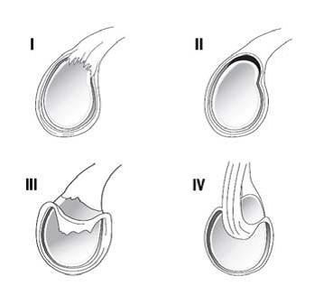
Figure 3: Synder’s four subtype classification system
Type I represents a frayed or degenerative labrum with attachment of the labrum to the glenoid. Type II represents superior labrum and biceps detachment from the glenoid rim. Type III represents a bucket-handle tear of the labrum with an intact biceps anchor. Finally, type IV represents a bucket handle tear of the labrum that extends into the biceps tendon. (From Dodson and Altchek 2009)
Synder et al. (1990) originally described and classified these tears into four distinct types (see figure 3)(12). This classification system later required some modifications. According to Maffet et al. (1995), only 62% of their shoulder series accurately fitted Snyder’s classification schema, so they composed a new classification system(11). As a result, they described an extra six new subtypes;
It is important to highlight that often, these SLAP lesions occur alongside other shoulder pathologies. It has been noticed that Type I lesions tend to occur alongside rotator cuff lesions, whereas type III and IV lesions tend to occur with traumatic shoulder instability. One study also showed that in type II lesions, older patients had rotator cuff injuries, whereas younger patients presented with shoulder instabilities(13).
Incidence
It has been recognized that acute SLAP tears are a relatively uncommon cause of shoulder pain and dysfunction in athletes taking part in overhead activities, such as tennis players, volleyball/water polo players, and baseball pitchers(2,13,14). While injury may occur in a one-off traumatic event such as a dislocated shoulder (where the humeral head impacts against the superior labrum and biceps anchor and creates a tear or a severe traction-type force to the biceps muscle), the majority are caused by repetitive wear and tear.
There are three potential overuse mechanisms underlying the pathophysiology of superior labral tears in overhead athletes(15,16):
1. Glenohumeral internal rotation deficit (GIRD) and internal impingement
It is known that glenohumeral external rotation increases over time in throwers (particularly baseball pitchers) due to the extreme rotation needed at late cocking and early acceleration of the throwing action. However, this is usually accompanied by a corresponding loss of internal rotation capacity due to tightness in the infraspinatus/ teres minor and posterior capsule of the shoulder seen on the throwing side of the body(14). This tight posterior shoulder musculature and posteroinferior capsule alter the GHJ kinematics so that the glenohumeral contact point shifts posterosuperiorly, especially during overhead-throwing activity(17). This creates an internal impingement of the articular side of the rotator cuff tendons and posterosuperior labrum between the humerus and the glenoid rim, precipitating a SLAP lesion(5). This internal impingement was first described by Walch et al. (1992) as an intra-articular impingement of the rotator cuff in the abducted and externally rotated shoulder(11).
2. Peel-back mechanism
During the late cocking phase of throwing, the biceps tendon is twisted at its anchor, which transmits torsional forces to the postero-superior labrum, resulting in a peel-back of the labrum(5,18,19). In a throwing shoulder, repeated initiation of this mechanism can lead to failure of the labrum over time with avulsion from the bone(18).
3. Eccentric biceps load
The biceps muscle acts eccentrically in the deceleration and follow-through stages of throwing. This eccentric load can place a traction effect on the superior labrum(7)
Injury Signs and Symptoms
The clinical diagnosis of a SLAP lesion can be an extremely challenging endeavor for the sports clinician because no unique clinical findings are associated with this type of pathology. Also, the condition is frequently associated with other shoulder problems such as impingement, rotator cuff tears, degenerative joint disease, and other soft tissue-related injuries(20). However, some common (but non-specific) features exist(21). These include:
- Dull, throbbing, ache in the joint.
- Difficulty sleeping due to shoulder discomfort. The SLAP lesion decreases the joint’s stability, which (when combined with lying in bed) causes the shoulder to drop.
- A catching feeling during throwing and pitching. Throwing athletes may also complain of a loss of strength or significantly decreased velocity in throwing.
Examination
There are numerous physical examination tests described to detect a SLAP injury. These can be sensitive but not very specific(5). These include:
1. Active compression / O’Briens’s Test
The shoulder is placed into approximately 90 degrees of forward flexion and 30 degrees of horizontal adduction across the body’s midline. Using an isometric hold, a downward resistance is applied in this position, with the entire shoulder’s internal and external rotation (altering humeral rotation against the glenoid in the process). A positive test for labral involvement is when pain occurs with the shoulder in internal rotation and forearm in pronation (thumb pointing toward the floor). Symptoms typically decrease when tested in the externally rotated position, or the pain is localized at the acromioclavicular (AC) joint. O’Brien found this maneuver to be 100% sensitive and 95% specific regarding assessing the presence of labral pathology(22).
2. Biceps load test II
During this test, the shoulder is placed in 90 degrees of abduction and is maximally externally rotated. At maximal external rotation and with the forearm supinated position, the patient is instructed to perform a biceps contraction against resistance. Deep pain within the shoulder during this contraction is indicative of a SLAP lesion. This test was further refined by describing the ‘biceps load II maneuver.’ The examination technique is similar, although the shoulder is placed at 120 degrees of abduction rather than the originally described 90 degrees. The biceps load II test was noted to have greater sensitivity than the original test. Only biceps load test II shows value in identifying patients with a SLAP-only lesion with no other concomitant pathology(23,24).
3. Uppercut test
This dynamic SLAP lesion test involves the patient rapidly bringing the hand up and toward the chin, replicating a boxing upper-cut punch. This upper-cut test had equal sensitivity (73%) and specificity (78%) in detecting a biceps tendon injury(25).
4. Compression rotation test
The compression rotation test is performed with the patient in the supine position. The glenohumeral joint is then compressed, and the humerus is rotated in an attempt to trap the labrum within the joint. The presence of an uncomfortable clunk may indicate a labral tear. The arm can be abducted with an anterior-directed force or adducted with a posterior-directed force to assess for anterior and posterior labral lesions.
5. Resisted supination external rotation test
Similar to the biceps load test, this test is performed with the patient in a supine and the shoulder at 90 degrees of shoulder abduction, with 65-70 degrees of elbow flexion and the forearm in a neutral position. The examiner resists against a maximal supination effort while passively externally rotating the shoulder(26).
6. Anterior slide test
This test is performed with the patient’s hands on the hips and the thumbs pointing posteriorly. One of the examiner’s hands is placed across the top of the shoulder from behind, while the other is positioned behind the elbow. A forward and slightly superiorly directed force is applied to the elbow and upper arm, and the patient is asked to push back against this force. Pain localized to the front of the shoulder under the examiner’s hand, pop or click in the same area, or both is considered a positive test(27). These tests form a cohort of tests that can all be used to attempt to assess for a SLAP lesion. While an individual test may not be overly sensitive, several positive tests would strongly hint at a SLAP lesion as the source of the shoulder pain. Another important part of the physical examination is the evaluation of scapular kinematics. There may be scapular dyskinesis, such as scapular winging, and poor retraction during the cocking phase of pitching. A postural analysis may show scapula downward rotation and anterior tilting. If a periscapular muscle atrophy or severe scapular winging is noted, an associating cervical spine pathology or accessory/long thoracic nerve palsy should not be excluded(28).
Imaging
Some authors believe that the only definitive way to detect a SLAP lesion is through arthroscopy and argue that imaging modalities will miss many of the subtle SLAP lesions as false negatives(29). MRI, particularly MR arthrography (MRA), is the gold standard imaging method to detect SLAP tears(30). The intra-articular injected contrast medium distends the joint capsule, outlines intraarticular structures, and leaks into tears(31,32). This, in turn, means a more precise delineation of the anatomic structures and SLAP lesions from anatomic variations like sub-labral recess or sub-labral foramen. A sub-labral recess or superior sulcus is a normal variant in more than 70% of individuals. In this variation, the base of the superior labrum is not attached to the superior glenoid; in some cases, this recess can be up to 1.4 centimeters deep(33).
MR arthrography can also detect spinoglenoid cysts. These cysts may cause entrapment of the suprascapular nerve, causing shoulder pain, weakness in external rotation, and infraspinatus muscle atrophy.
SLAP tears are best seen on coronal oblique sequences with the arm abducted and externally rotated, as the contrast medium fills the gap between the glenoid and superior labrum(34). Although MR arthrography is the best imaging technique to evaluate SLAP lesions, a high incidence of false positives is possible; therefore, findings must be correlated with clinical history and physical examination.
Summary
A SLAP lesion is an uncommon shoulder pathology, affecting primarily athletes involved in an overhead sport. Baseball pitchers seem to be the most vulnerable group due to the unique forces they place on the biceps-labral complex in pitching. Part one of this Rehabilitation Masterclass on SLAP lesions was to present the relevant anatomy and pathomechanics of the shoulder about SLAP lesions and how a SLAP lesion is most often diagnosed. Part two will discuss in detail the surgical management of a SLAP lesion and, in particular, how it is rehabilitated in the event of shoulder surgery to repair the biceps anchor.
References
-
Am J Sports Med 2013; 41: 1372-1379
-
J Shoulder Elbow Surg. 1995;4:243-248
-
J Shoulder Elbow Surg 2010; 19: 1199-1203
-
J Bone Joint Surg Am 1992; 74: 46-52
-
Am J Sports Med 2013; 41: 444-460
-
J Bone Joint Surg Am. 1995;77:1003-1010
-
Am J Sports Med. 1985;13(5):337-41
-
Arthroscopy 1990; 6: 274-279
-
Am J Sports Med 2012; 40: 1538-1543
-
Journal of orthopaedic & sports physical therapy. 2009; 39(2), 71-80
-
Am J Sports Med 1995; 23: 93-98
-
Arthroscopy 1990; 6: 274-279J Am Acad Orthop Surg. 1998;6:121-131.
-
Am J Sports Med 2011; 39: 1687-1696
-
Am J Sports Med 2012; 40: 2536-2541
-
Arthroscopy 2003; 19: 641-661
-
Int J Sports Phys. 2013; 8(5). Pp 579-600
-
Arthroscopy 2003; 19: 404-420
-
Arthroscopy 1998; 14: 637-640
-
Am J Sports Med. 2001;29:488-492
-
Oper Tech Sports Med 2004; 12: 99-110
-
Eur J Radiol. 2008; 68 (1): 72–87
-
Am J Sports Med. 1998;26(5):610-3
-
Clin J Sport Med 2010; 20: 134-135
-
J Shoulder Elbow Surg 2012; 21: 13-22
-
J Sports Med. 2009;37(9):1840–1847
-
Am J Sports Med. 2005;33:1315-1320
-
Arthroscopy. 1995; 11:296-300
-
J Athl Train. 2011;46(4):343–348
-
Am J Sports Med. 1994;22(4):493-8
-
Radiology. 2000;214(1):267-71
-
Radiology 2001; 218: 127-132
-
AJR Am J Roentgenol 1993; 161: 1229-1235
-
Arthroscopy 1998; 14: 789-796
-
Clin Sports Med 2008; 27: 607-630
Further reading
Newsletter Sign Up
Subscriber Testimonials
Dr. Alexandra Fandetti-Robin, Back & Body Chiropractic
Elspeth Cowell MSCh DpodM SRCh HCPC reg
William Hunter, Nuffield Health
Newsletter Sign Up
Coaches Testimonials
Dr. Alexandra Fandetti-Robin, Back & Body Chiropractic
Elspeth Cowell MSCh DpodM SRCh HCPC reg
William Hunter, Nuffield Health
Be at the leading edge of sports injury management
Our international team of qualified experts (see above) spend hours poring over scores of technical journals and medical papers that even the most interested professionals don't have time to read.
For 17 years, we've helped hard-working physiotherapists and sports professionals like you, overwhelmed by the vast amount of new research, bring science to their treatment. Sports Injury Bulletin is the ideal resource for practitioners too busy to cull through all the monthly journals to find meaningful and applicable studies.
*includes 3 coaching manuals
Get Inspired
All the latest techniques and approaches
Sports Injury Bulletin brings together a worldwide panel of experts – including physiotherapists, doctors, researchers and sports scientists. Together we deliver everything you need to help your clients avoid – or recover as quickly as possible from – injuries.
We strip away the scientific jargon and deliver you easy-to-follow training exercises, nutrition tips, psychological strategies and recovery programmes and exercises in plain English.
