You are viewing 1 of your 1 free articles
Thoracic Outlet Syndrome in the Athlete: Part 2
Chris Mallac outlines the clinical tests used to diagnose TOS and discusses conservative management and surgical options to treat this injury.
Clinical Tests
There are several clinical tests purported to diagnose TOS. However, many of these tests have high false-positive rates (up to 50%)(1). A cluster of several positive tests may improve their sensitivity and reliability(2).
These tests include;
- Roos Test or Elevated Arm Stress Test (EAST)(3)
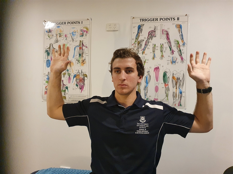
- Athlete raises the arm to 90 degrees shoulder abduction, external rotation, and 90 degrees elbow flexion (‘High 5’ position) (see figure 1).
- The athlete then opens and closes the hands for up to 3 minutes to reproduce symptoms. Most patients with neurogenic TOS will experience symptoms within 20-30 seconds, and most cannot continue past 1 minute.
- Reproduction of symptoms may indicate compression in possibly all three compression areas (scalenes, costoclavicular or axillary)(4)
- Sensitivity of this test is 52%-84%, meaning the test will detect 52% to 84% of all positive cases. At the same time, the specificity is 30% to 100%, meaning that the likelihood of a positive test for TOS that is another pathology is 30% to 100%(2,5,6).
- Supraclavicular Pressure
- Patient sits in a relaxed position.
- Examiner places fingers on upper trapezius and thumb on anterior scalene.
- Examiner squeezes thumb and fingers together.
- Pain or paraesthesia reproduction represents a positive test
- Specificity 85% to 98%(6).
- Adson’s Test
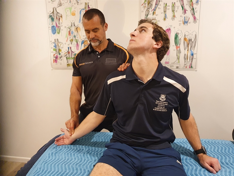
- The clinician raises the arm into some elevation, slight external rotation, slight extension, and feels the pulse (see figure 2).
- The athlete extends and rotates the head to the affected side.
- The athlete takes a deep breath and holds the breath.
- A diminished pulse or paraesthesia may indicate compression between the anterior and middle scalene.
- Sensitivity of 79% and specificity of 74% to 100%(2,6).
Related Files
- Eden’s Test or Military Brace Test
- The clinician raises the arm into slight external rotation, slight extension and feels the pulse.
- The clinician then tractions the arm by pulling on it and applies gentle pressure to the clavicle to depress it.
- A diminished pulse or reproduction of symptoms may indicate compression in the costoclavicular space.
“A cluster of several positive tests may improve their sensitivity and reliability(2).”
- Costoclavicular Sign
- The athlete draws the shoulders into retraction and depression.
- The clinician feels for the pulse.
- A diminished pulse may indicate compression between the clavicle and first rib or between the subclavius or costocoracoid ligament.
- Specificity of 53% to 100%(6).
- Wrights Test or Hyperabduction Test
- Clinician takes arm into abduction, extension, and external rotation.
- The clinician feels for a pulse.
- The athlete then takes a deep breath, holds it, then rotates the head.
- An alteration in pulse may indicate a compression under the pectoralis tendon.
- Sensitivity of 70% to 90% and specificity of 29% to 53%(6).
- Upper Limb Tension Test 1
- Athlete lies supine with their arm at 90 degrees of abduction and external rotation.
- Clinician moves arm into elbow extension and wrist extension.
- A reproduction of symptoms indicates a positive neurogenic TOS.
- Sensitivity of 90% and specificity of 38%(6).
Correcting scapular postures during these tests may also indicate the importance of scapular posture in the pathophysiology of TOS. If scapular repositioning during any of the tests changes the symptoms, the clinician should address scapular posture during rehabilitation(4).
Imaging
In most cases, diagnostic procedures such as doppler ultrasound, electromyography, and nerve conduction studies are non-conclusive and ineffective in diagnosing neurogenic TOS(7). However, precise nerve conduction studies may identify differences in median nerve versus medial antebrachial cutaneous nerve and ulnar nerve sensory responses. Determining specific nerve involvement may help the clinician perform a differential diagnosis that distinguishes TOS from other pathologies, such as cervical radiculopathy(8,9).
X-ray studies are only helpful to identify whether the patient is suffering from a definitive structural abnormality at the first rib, the presence of a cervical rib, or a grossly malunited previous clavicle fracture. Xray may show a widening of the first rib at the vertebral or sternal attachment(10).
Magnetic Resonance Imaging (MRI) may show abnormalities in the brachial plexus; however, this adds little value in diagnosing symptomatic TOS(8). An MRI may prove more useful in the rarer versions of vascular TOS. Magnetic Resonance Angiography and Magnetic Resonance Venography allow injection of blood pooling agents (BPA) and extracellular contrast agents (ECA), which may help identify fixed structural abnormalities or functional blockages of the blood vessels(11). Furthermore, Magnetic Resonance Neurography (MRN) can identify brachial plexus compression from fibrous bands in and around the interscalene triangle(12).
“If scapular repositioning during any of the tests changes the symptoms, the clinician should address scapular posture during rehabilitation(4).”
Treatment
The more common variation of neurological TOS – the symptomatic TOS – is devoid of a structural neurological or vascular lesion. Therefore, the first option for management is conservative treatment for four to six months. Consider surgery only in failed and recalcitrant cases. Burt (2018)(13) at the Baylor College of Medicine created a treatment algorithm that suggests that cases of neurological TOS that show less than 25% improvement after four weeks of targeted physiotherapy are likely surgical candidates. However, a study conducted at Vanderbilt University showed that 59% of the 42 patients with neurological TOS had symptomatic relief after six months of conservative physiotherapy(14).
A host of mitigating factors determines the timing of surgical intervention in the athlete. Some considerations include the competition training phase, the time needed away from sport for rehabilitation, and resulting changes to ergonomics and technique that may impact performance.
Conservative Treatment
Initial conservative treatment starts with the following measures:
- Relative rest depending on how provocative the symptoms are with activity.
- A short course of NSAIDs to alleviate provoked symptoms.
- First rib mobilization and scalene stretches and releases.
- Nerve mobility exercises.
- Botox injections into the scalenes if they are the cause of the symptoms. A study at UCLA found that botulinum toxin injections relieved pain, numbness, and fatigue by up to 50% for one month in 64% of the subjects(15). However, a Cochrane Review showed moderate evidence that Botox injections into the scalenes did not improve pain and disability(16), although this treatment approach could improve neurological symptoms, such as paraesthesia.
- Manage other concurrent pathologies that may complicate the TOS, such as subacromial bursitis, shoulder instability, or rotator cuff pathology.
- Address scapular muscle imbalances and dysfunction.
Scapular Dysfunction
Typically, patients who suffer from TOS demonstrate scapular dysfunction(17). The scapula is usually depressed or downwardly rotated and referred to as the ‘dropped shoulder condition’(17). This scapular posture leads to a decrease in the anatomical space between the clavicle and first rib. Thus, this position reduces the costoclavicular space and compresses or stretches the neurovascular bundle.
The athlete may demonstrate the dropped scapula posture at rest and during functional arm movements. The resulting scapular dyskinesis manifests as late and insufficient upward rotation with shoulder abduction, an anterior tilt with shoulder flexion, or a ‘winging’ scapula with the above movements.
Both weakness and lack of neuromuscular control in the serratus anterior and upper, middle, and lower trapezius muscles contribute to scapular dyskinesis. A dominance and over-reliance on the rhomboids, pectoralis minor, and levator scapulae also add to the abnormal movement patterns(17). If manually repositioning the scapula changes the outcome of a provocation test, the scapular dyskinesis may be the main culprit in the TOS symptoms(17).
Several interventions can correct the dropped scapula posture:
- Stretch and perform manual therapy to the pectoralis minor, rhomboids, and latissimus dorsi to limit depression and downward rotation.
- Use strapping tape to fashion an ‘axillary sling’ to hold the scapula into more elevation and upward rotation(17).
- Strengthen the upward rotators with isometric holds in neutral positions and progress to more active upward rotation in elevated arm positions(17). The reader is directed to the rehabilitation ideas proposed by Watson et al.(2010)(17).
- Amend postures in activities of daily living to limit cervical flexion and arm abduction. Practice belly breathing to offload accessory respiratory muscles.
Surgical intervention
The purpose of surgery in TOS management is to remove any extrinsic compression on the neurovascular bundle within the thoracic outlet. Vascular TOS (arterial or venous) usually requires surgery as a structural anomaly is almost always present (such as a cervical rib, hypertrophied scalenes, or large first rib). If a structural lesion is present, conservative management will fail, and the patient will invariably require surgery to remove the occlusive defect.
It is often difficult to detect a structural anomaly in the case of venous occlusion. Venous TOS treatment often consists of a catheter-directed thrombolytic injection, followed by anticoagulation for three months before surgery(18). The patient usually requires additional post-surgical anticoagulation with repeated venograms to ensure the subclavian vein regains patent(18,19, 20, 21).
Arterial TOS requires surgical decompression by removing bony abnormalities. The patient may require reconstruction of the arterial lesions using bypass grafts between the proximal subclavian artery and axillary artery(22).
In cases of the more common neurological TOS presentation, a trial period of conservative treatment is attempted before surgery, as conservative management of neurological TOS can be successful. In a study of Major League Baseball pitchers who underwent surgical decompression for neurological TOS, the majority had excellent outcomes in pitching metrics such as velocity, movement, spin rate, and spin direction. The authors highlight that with a lengthy post-operative rehabilitation period (approximately 11 months), elite-level pitchers can return to as good as, if not better, pitching metrics as they enjoyed before surgery(23).
Some of the more common surgical options are(7,13,16,):
- First rib removal may be performed via a trans-axilla approach, a supraclavicular approach, or an infraclavicular approach.
- Scalene resection, particularly anterior scalenectomy(10).
- Combined 1st rib resection and scalenectomy (FRRS).
- Brachial plexus neurolysis.
- Pectoralis tendon tenotomy.
“The purpose of surgery in TOS management is to remove any extrinsic compression on the neurovascular bundle within the thoracic outlet.”
Two primary incisions may be used – a supraclavicular or a trans-axillary. The supraclavicular approach is the preferred option as it allows complete removal of the scalenes, visualization of anatomical variants, and better access to the brachial plexus for neurolysis(7). A Cochrane Review in 2014 (Povlsen 2014) found low-level quality evidence that trans-axillary first rib resection (TFRR) is slightly superior to supraclavicular neurolysis of the brachial plexus (SNBP) for pain relief in patients who failed conservative treatment(16).
Conclusion
Thoracic outlet syndrome is a poorly defined condition with a host of signs and symptoms overlapping with other differential pathologies. The clinical diagnostic tests have a high false-positive rate, and imaging is often unhelpful, particularly in neurological TOS.
If identified, vascular TOS usually requires more formal surgical or anticoagulation interventions. The more common neurological TOS may respond to conservative management that focuses on improving scapular muscle control, positioning, and movement.
References
- Current Sports Medicine Reports. 2014, 13(2); 81-85.
- Joint Bone Spine. 2001; 68(5): 416-424.
- J Vasc Surg. 2017;66(2):533–44
- International Journal of Osteopathic Medicine, 2010; 133e142
- Journal of Manual & Manipulative Therapy, 2010;18:2, 74-83
- Journal of Manual & Manipulative Therapy, 2010; 18:3, 132-138
- Current Reviews in Musculoskeletal Medicine, 2020; 13:457–471
- Hand Clinic. 2004; 20: 27-36.
- Muscle Nerve. 2014; 49(5): 724-7.
- Ann Vasc Surg. 2007; 21: 564-570.
- Eur J Radiol. 2014; 83:1209-1215.
- Eur Radiol. 2014; 24:756-761.
- J Thoracic Cardiovasc Surg. 2018; 156(3). 1318-1323.
- J Am Acad Orthop Surg. 2015; 23(4): 222-32
- Vasc Surg. 2000;14:365–9.
- Cochrane Database Syst Rev2014 Nov 26;(11):CD007218.
- Manual Therapy, 2010.15(4); 305-314
- Arch Surg. 1989, 124: 1153-1158.
- 2009, 145: 500-507.
- J Vasc Surg 2001;35:181e97.
- J Vasc Surg. 2017; 65(5); 1429-1439.
- Current Opinion in Cardiology. 2010. 25: 535-540.
- Ann Vasc Surg 2017; 39: 216–227Pain Therapy. 2019, 8: 5-18
Newsletter Sign Up
Subscriber Testimonials
Dr. Alexandra Fandetti-Robin, Back & Body Chiropractic
Elspeth Cowell MSCh DpodM SRCh HCPC reg
William Hunter, Nuffield Health
Newsletter Sign Up
Coaches Testimonials
Dr. Alexandra Fandetti-Robin, Back & Body Chiropractic
Elspeth Cowell MSCh DpodM SRCh HCPC reg
William Hunter, Nuffield Health
Be at the leading edge of sports injury management
Our international team of qualified experts (see above) spend hours poring over scores of technical journals and medical papers that even the most interested professionals don't have time to read.
For 17 years, we've helped hard-working physiotherapists and sports professionals like you, overwhelmed by the vast amount of new research, bring science to their treatment. Sports Injury Bulletin is the ideal resource for practitioners too busy to cull through all the monthly journals to find meaningful and applicable studies.
*includes 3 coaching manuals
Get Inspired
All the latest techniques and approaches
Sports Injury Bulletin brings together a worldwide panel of experts – including physiotherapists, doctors, researchers and sports scientists. Together we deliver everything you need to help your clients avoid – or recover as quickly as possible from – injuries.
We strip away the scientific jargon and deliver you easy-to-follow training exercises, nutrition tips, psychological strategies and recovery programmes and exercises in plain English.
