You are viewing 1 of your 1 free articles
The long head of the biceps tendon - Part I
In part one of this two-part article, Chris Mallac explores the anatomy and function of the long head of the biceps tendon, and the types of injuries sustained
Injuries to the long head of the biceps tendon (LHBT) are reasonably common injuries in throwing athletes and athletes in sports with repetitive hand-above-head positions. These include swimmers, tennis players, cross-fit athletes and gymnasts. Injuries can be as simple as a tendinitis or tenosynovitis, and extend to more traumatic injuries such as full ruptures.
The anatomy of the LHBT and its corresponding structures has been extremely well researched over the last century. You might therefore expect that the function of this structure and its role in shoulder injury is well understood. However, conjecture remains over the exact anatomy and function of this unique anatomical tissue.
Relevant anatomy
The LHBT is unique as it has both an intra-articular structure that originates inside the glenohumeral joint, becoming extra-synovial as it passes through the anterior rotator interval to enter the bicipital groove. The proximal long head tendon is approximately 9cm long and 5 to 6mm in diameter. The articular portion is flatter and a little larger than the groove portion, which is round and smaller and has an oblique inclination of 30-40 degrees. The tendon then runs through intertubercular groove and courses further down into the bicipital groove, protected by a synovial sheath(1).
In terms of its anatomy and function, LHBT may be likened to the components of a boat:
- The ‘anchor’ is the proximal attachment of the tendon onto the supraglenoid tubercle and labrum (approximately 50% originate on the tubercle and 50% onto the labrum)(2).
- The ‘pulley’ is the tendon component that runs over the humeral head and is supported by the ligaments that create the ‘biceps reflection pulley system’ (discussed in next section).
- The ‘gunwale’ is formed by the hard edges of the humerus bone, which form the bicipital groove and houses the tendon and its corresponding sheath as it courses down the humerus to join the biceps muscle.
The ‘biceps reflection pulley system’
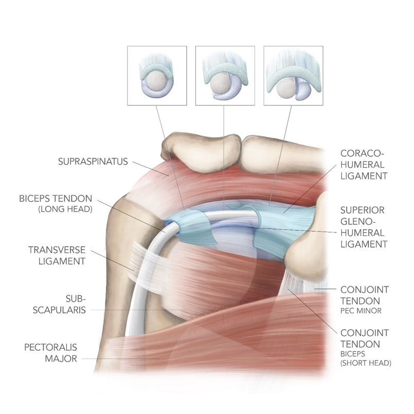
Proximal to the bicipital groove, the tendon is stabilised by a ‘biceps reflection pulley system’ (see figure 1). The superior glenohumeral ligament (SGHL) and the coracohumeral ligament (CHL) are the structures that envelope the tendon, and which make up the pulley system - with the SGHL being the more important stabiliser of the LHBT(3).
Furthermore, in the bicipital groove, the tendon is stabilised by the transverse humeral ligament, which is formed by fibres from the subscapularis and supraspinatus tendons(4). However, the transverse humeral ligament is not a significant stabiliser of the ‘biceps pulley’ at the entry of the groove. Doubt has been expressed about the existence of the transverse ligament as a distinct entity, and it may simply be a continuation of the insertion of subscapularis(4). Furthermore, the pectoralis major tendon also crosses over the tendon in the bicipital groove.
Figure 1: Anatomy of the LHBT showing the biceps reflection pulley system

Interestingly, a number of proximal variants have been described to the LHBT and this has led to some confusion amongst radiologists and surgeons performing arthroscopic investigations(2,5-8). These include:
- Congenital absence of the tendon.
- A synovial ‘mesentery’.
- Adherence to the supraspinatus tendon and fusion with rotator cuff(9).
- Bifurcated origin tendons.
- Extra-articular segment.
- Presence of a vincula (band of connective tissue).
The LHBT is vascularised by branches of the suprascapular artery, the anterior humeral circumflex artery and the deep brachial artery(10). The blood supply to the tendon has been described in two anatomical zones – the traction zone and the sliding zone(11). A characteristic vascular pattern is seen on the superficial surface of the tendon within the groove (traction zone), but the deep ‘sliding’ surface is avascular and composed of fibrocartilage. There is a consistent hypovascular area 1 to 3cm from the tendon origin, possibly explaining the susceptibility of this area to rupture(11, 12).
Function of the LHBT
Despite anatomical, biomechanical and electromyographic studies, there remains a lot of controversy about the function of the LHBT at the shoulder. Overall, the primary functions attributed to the LHBT have been humeral head depression, glenohumeral stabilisation and external rotation of the shoulder. To summarise, below are some key studies that have looked at the function of the LHBT:
- The long head of the bicep is a weak abductor of the shoulder (only 7% to 10% of total action)(13).
- Contraction of the biceps, or external rotation of the arm, provides stability to the humeral head, preventing superior migration of the humeral head(14-16).
- Electromyographic testing did not show muscle activity in the bicep with external rotation when the elbow was kept immobilised. This may implicate its function is dependent on elbow movement(17-18).
- In patients with rotator cuff tears, increased biceps activity was shown. This suggests that it may act in a compensatory way to provide shoulder stability in a shoulder with cuff damage(16).
- Detachment of the superior labrum and biceps anchor causes an increased anterior and inferior translation of the humeral head on the glenohumeral joint, with more tension transmitted to the inferior glenohumeral ligament in the cocking position of throwing(19, 20). This suggests that the LHBT has a role in anterior shoulder stability when the bicep contracts with the throwing action.
- In other throwing-based studies, it was shown that in an unstable shoulder, the contribution to anterior stabilisation is increased, with greater electromyographic activity of the biceps muscle in such individuals during throwing motion(21-24).
As the human structure has evolved over time, the scapula has moved into a more frontal plane with an associated torsion of the humerus, thus reducing the action of the LHB at the shoulder(25,26). Due to the torsion through the humerus, the bicipital groove is no longer centred on the plane of the humeral head, but lying at an angle of about 30 degrees to it(27). This creates a pulley system with the lesser tuberosity of the humerus, and as a consequence the LHBT is forced against the lesser tuberosity and the medial wall of the groove, instead of at the middle of the groove. This pulley action of the medial wall and lesser tuberosity renders the tendon vulnerable to trauma. In fact, over time, due the changing structure and orientation of the scapula and humerus to suit modern day human function, it is now believed that the LHBT is a vestigial structure that is no longer functionally needed(17, 18).
Pathology in the LHBT
Pain arising from the LHBT can stem from either the intra-articular portion due to inflammation, instability and rupture, and/or the extra-articular portion in the bicipital groove, which may be prone to injury due to its close association with the tendon sheath. Being both intra-articular and extra-synovial, places unique forces on the LHBT during movement, and this may lead to particular injury patterns.
Being an intra-articular but extra-synovial structure ensures it is essentially static within the joint. It slides passively on the humeral head during abduction or rotation, and this creates internal shear across the tendon and bone(25). Furthermore, due to its anatomical location within the shoulder, the LHBT may also be subjected to extraarticular impingement in the subacromial space.
Disorders of the biceps tendon can be classified into degenerative, inflammatory, mechanical/instability and traumatic (rupture). However, different pathologies usually co-exist. Although isolated pathology of the biceps does exist, it has a high association with rotator cuff tears (particularly supraspinatus) and is also associated with abnormalities of the glenoid labrum. For the purposes of this article, the discussion will focus on tendon lesions at biceps pulley and in the bicipital groove and its association with rotator cuff pathology. Superior glenoid attachment injuries such as SLAP lesions will not be discussed as they have been discussed in previous Sports Injury Bulletin editions (see issues 135, 155 and 156).
Mechanisms of injury
The typical mechanisms of injury to the pulley system can include:(28-30)
- A fall on the outstretched arm in combination with full external or internal rotation.
- A fall backward on the hand or elbow.
- A forcefully stopped overhead throwing motion.
- During throwing actions, with active contraction of the biceps in internal rotation, the strain increases in the bicep whilst the elbow is decelerated in extension. By this deceleration, a maximal contraction of the LHB is provoked, which can cause tears in the rotator interval capsule.
- Repetitive, forceful internal rotation above the horizontal plane. This causes friction damage between the pulley system and subscapularis on the one hand, and the anterior superior glenoid rim on the other.
Ruptures of the LHBT are more commonly associated with strong biceps contraction whilst in shoulder external rotation, such as making a tackle in football. The combined tensile and torsional force may breach the tensile load failure point of the tendon, particularly in the presence of a degenerative tendon.
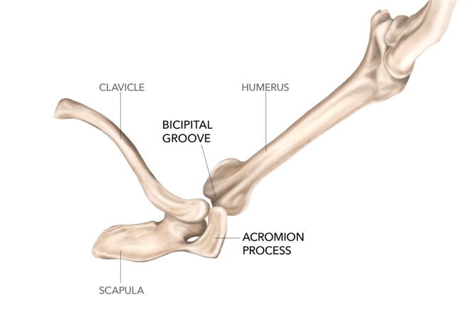
Other injuries that cause inflammation, and subsequent long-term degeneration include impingement of the LHBT. This may be due to the close approximation of the bicipital groove (and hence LHBT) and the anterior aspect of the acromian process when the arm is fully elevated overhead (see figure 2). This would be a potential provocative movement in overhead athletes who repeatedly use their arms above the head such as swimmers, cross fit athletes and tennis players.
Figure 2: The potential impingement of the bicipital groove onto the acromian process

Shoulder injuries in swimmers and cross-fit athletes
Shoulder pain is the most common debilitating syndrome that affects freestyle/butterfly swimmers. McMaster has shown that there exists a prevalence of 35% of shoulder pain in competitive swimmers(31). Becker has also suggested that female swimmers will most likely suffer shoulder pain at least three times in their swimming career(32). The following discussion is designed for a typical North American swimming programme; however the discussion can be extrapolated for any swimming population, irrespective of the country of residence:
- During mid adolescence (as the bodyweight increases but the muscle system has still not fully developed) the mass increase and the resultant increase in drag in the water creates an overload situation in the shoulder.
- At the end of adolescence (during the later stages of high school), the body has reached maximum body weight. However, it is still not muscularly strong enough to withstand the stressors from the harder training.
- The third period is the transition from high school to college where swim training volume increases considerably. However, freshman swimmers are often afraid to report initial onsets of shoulder pain due to the fear of falling behind with their swim programme.
Male swimmers however tend to have two peak times of shoulder pain:
- The end of the second growth spurt in adolescence, when body weight increases but muscle strength has not yet caught up to the increase in bodyweight.
- The second peak time is in the first college year, when around December time, the training load suddenly increases over a short time period.
‘Swimmers shoulder’, which involves a subluxation of the shoulder that impinges the long head of the bicep and the supraspinatus tendon, was first described in 1978 by Kennedy and Hawkins(33). The reach position at the catch point of freestyle involves maximum shoulder flexion/abduction. At this point the hand is attempting to forcefully supinate whilst the arm is aggressively pulled through the water with elbow flexion. These movements both require forceful biceps contraction. Whilst this is happening, the arm is forcefully internally rotating, creating an anterior shear of the humeral head in the glenoid socket. As the shoulder pushes and rolls forward, the biceps tendon is subjected to an added strain.
The risk of impingement here is the greatest, and the stress on the biceps tendon and rotator cuff tendons is the greatest. Therefore, impingements syndromes involving the bursa, supraspinatus tendon and biceps tendon and tendinopathies to the rotator cuff tendons are a significant injury risk.
These unique forces on the shoulder are attributed as being the damaging forces on the soft tissue structures in and around the shoulder. The swimmer needs not only adequate flexibility in shoulder rotation, but also flexibility in the thoracic spine to allow the reach to occur. Furthermore, the shoulder needs adequate strength in the rotation control muscles – the rotator cuff - as well as well-conditioned scapular stabilisers and mobilisers.
Cross-fit athletes are of particular interest due to the unique skills they need to perform particular workouts. Movements such as ‘handstand walks’, ‘handstand push ups’, ‘kipping chins’ and ‘overhead snatch grip squats’ may all be potentially impinging positions (see figure 3). In a survey based study on shoulder injuries in cross fit, it was found that gymnastics based movements and Olympic based movements seemed to be the most provocative loads on the shoulder for athletes(34).
Figure 3: Cross-fit athlete in a potentially impinging position

Types of injuries to the LHBT
*Biceps tendinopathy
The terms tendonitis and tenosynovitis are commonly used to describe irritation of the tendon and its sheath within the bicipital groove. Although the understanding of degenerative tendinosis has made rapid progress more recently(35), relatively little work has focused on the rotator cuff, and none specifically on the LHBT. Therefore, ideas on tendinosis and tendon pathology need to be extrapolated from other more well-researched tendons such as the Achilles and patella(35). Tenosynovitis, tendinosis, delamination, pre-rupture and rupture probably represent the natural history of progressive degeneration of the biceps.
Due to its anatomy, with a synovial sheath and constrained path in the bicipital groove, the LHBT is subject to tendonitis/tenosynovitis(36). Pain in and around the tendon sheath may represent a chronic ‘stenosing’ degenerative process similar to the pathology that affects the first dorsal compartment of the wrist (known as deQuervain’s syndrome)(37).
With regards to the LHBT, it may be caused by the following:
- Attrition of the tendon and sheath in the groove due to abnormalities such as osteophytes under the tendon.
- A compressive transverse humeral ligament.
- A shallow and narrow shape of the groove, which increases the compressive force across the tendon and sheath(36, 38).
- Patho-mechanical faults due to scapula dyskinesis (muscle imbalance issues in and around the scapula and thoracic spine).
However, it is believed that primary tendonitis is an uncommon pathology occurring in only approximately 5% of all cases of biceps tendinopathy, and if this does occur it is more likely in younger throwing athletes, or athletes in repetitive hand above head postures(39). It is more likely that it is present along with rotator cuff pathology. The majority of degenerative changes in the LHBT are associated with pathology of the rotator cuff(40, 41).
To support this idea, in a study of complete rotator cuff tears, Chen et al found that 76% of cuff tears had associated LHBT pathology(42). Gill et al showed that in 85% of LHBT partial tears, cuff pathology was associated(43). As the degeneration evolves, the tendon may fibrillate and then split, and hypertrophy or attenuation may occur. This may be described as delamination or pre-rupture.
To explain the association between LHBT injury and rotator cuff damage, a patho-mechanical model was proposed by Refior and Sowa(44). This suggested that an upward migration of the humeral head due to rotator cuff damage leads to repetitive traction, friction, and glenohumeral rotation. Pressure and shear forces can occur on the tendon at distinct, anatomically narrow sites, resulting in degenerative changes such as fibrosis, thickening, collagen disorganisation, scar tissue, and development of adhesions.
*Biceps instability
The unique anatomy of the LHBT (with its pulley system) is responsible for the stability of the tendon as it courses from the intra-articular space into the bicipital groove. The pulley is formed by four structures (see anatomy above)(30):
- The coracohumeral ligament (CHL).
- The superior glenohumeral ligament (SGHL).
- Fibres from the subscapularis tendon.
- The supraspinatus tendon.
An injury to the pulley system can be secondary to a traumatic event, which damages the supporting ligament structures, or to a degenerative process that affects the supraspinatus and/or subscapularis (see box 1) (28,45). In the event of a torn pulley system, the LHBT becomes unstable. As it becomes unstable it may displace and sublux or dislocate from the bicipital groove.
| Type 1 | Isolated injury to the SGHL. |
| Type 2 | SGHL lesion and supraspinatus tendon lesion. |
| Type 3 | SGHL lesion and subscapularis tendon lesion. |
| Type 4 | Lesions of all the structures. |
*LHBT subluxation
A subluxation of the LHBT is a partial and/or transient loss of contact between the tendon and its groove. This creates pain with no sensation of locking or loss of function. A dislocation is the complete and permanent loss of contact between the tendon and the groove. In a dislocation, patients may suffer a ‘pseudoparalysis’ of the shoulder because of the associated rotator cuff pathology(46).
*LHBT dislocation
Dislocations of the LHBT can be classified into intra-articular, intra-tendinous, and extra-articular subtypes. Dislocation may be associated with a tear of subscapularis tendon or (where the subscapularis remains intact) when the LHBT dislocates over or under the subscapularis tendon(46, 47). A LHBT dislocation with an intact subscapularis tendon implies damage of the rotator interval tissue, including the CHL and SGHL (48). Biceps tendon dislocations medial to the lesser tuberosity are usually accompanied by tears or attenuation of the ligamentous pulley(46). The transverse ligament overlying the bicipital groove is not considered a crucial stabilising structure unless the medial CHL is torn(49).
Supraspinatus pathology is commonly associated with LHBT lesions, and this may have implications for the stability of the LHBT in the groove. At its posterior edge, the supraspinatus is the restraint to movement of the LHBT. Damage to the supraspinatus and thus the superior border of the pulley may lead to subluxation and eventually dislocation of the LHBT.
Dislocation may also produce a contour change in the biceps muscle due to shortening of the course of the tendon. This has been referred to as the ‘hourglass biceps’. The result is that the tendon becomes hypertrophic - often in association with advanced disease of the rotator cuff - and is unable to slide into the bicipital groove. This presentation is more common than the tendon becoming fixed in the bicipital groove by adhesions(25). Both have the same mechanical effect of buckling of the tendon on elevation of the shoulder, with entrapment of the tendon between the humeral head and glenoid. This leads to pain and a block to terminal elevation.
*LHBT instability
Instability of the LHBT is a common injury pattern in throwing athletes due to the high prevalence of SLAP lesions found in athletes(50). In throwing athletes, the contact of the pulley with the posterosuperior labrum in the late cocking phase can damage the pulley(51). Bennett et al found that in 43% of SLAP repairs there was pulley system damage(52).
The more common variation of LHBT instability is medial instability as discussed above. Lateral instability may also happen, although this is rare. This has been described mainly in a traumatic context following anterior shoulder dislocation and/or fractures of the greater tuberosity(53,54). However, posterior and lateral instability can also be found in association with supraspinatus tears. Dynamic exploration under arthroscopy or open surgery shows that, in the presence of a supraspinatus tear, the LHB can roll over the lateral rim of the groove when the arm is placed in abduction and internal rotation.
Tendon ruptures
Similar to other ruptures of tendons, LHBT ruptures are commonly secondary to a degenerative process, which may be due to tendon instability and/or impingement syndromes, and usually occur in the presence of a rotator cuff tear. The usual mechanism of injury in the athlete will be a forced bicep contraction in a stretch position (such as making a rugby tackle), but it may also happen in innocuous activities of daily living.
Rupture of the LHBT will usually create a deformity in the contour of the biceps muscle due to the distal migration of the long head of the bicep and this is commonly called a ‘Popeye sign’. However, in some cases, the presence of a vincula, adhesion or hypertrophy of the tendon can prevent distal migration of the tendon and the subsequent ‘Popeye sign’(55). Tendons that dislocate often become encased in fibrous tissue or adherent to the subscapularis before rupture, and hypertrophic tendons may become fixed within the bicipital groove, giving an autotenodesis. If the rupture does occur in the tendon substance in the biciptal groove (and the distal end retracts and creates the ‘Popeye sign’), the proximal end or stump may remain with the joint, and cause pain as it is compressed between the humeral head and the glenoid(56, 57).
Conclusion
The LHBT is a unique anatomical structure within the shoulder joint complex. It has a complex interplay with the ligaments of the shoulder, which create the ‘biceps reflection pulley system’. However, its functional role in shoulder movement and stability has been questioned. It has been suggested by some authors as being a weak abductor and external rotator of the shoulder, and its primary role as being a glenohumeral stabiliser. Others have refuted this idea and claim it has become a redundant structure, similar to the appendix. However, it can be damaged in athletes that participate in sports that require repetitive biceps contraction in vulnerable positions or due to compressive forces whilst the shoulder is constantly elevated. The injuries can range from simple tendinitis and tenosynovitis, through to damage to the ‘pulley system’ and finally rupture of the LHBT. In part two, we will describe how LHBT injuries can be diagnosed and their management.
References
- J of Bone Joint Surg. 2007; 89-B (8), 1001-1009
- J Bone Joint Surg Br. 1994; 76:951–4
- Am J Sports Med 2000;28:28-31
- Am J Sports Med 2006;34:72–7.
- Unfall chirurg. 1987;90:319–29
- Arthroscopy 2004;20:1081–3
- J Shoulder Elbow Surg 2007;16:e25–30
- J Shoulder Elbow Surg 2008;17:114S–17S
- Rev Bras Ortop. 2016. 51(1); 96-99
- Burkhead W. The biceps tendon. In: Rockwood CJ, ed. The shoulder. Philadelphia, Pennsylvania, USA: WB Saunders, 2004:1059–150
- Orthopaedics 2006;29:149-52
- Clinical Anatomy. 2010; 23(6), 683–692
- Acta Anat (Basel). 1976; 96:270-84
- Clin Orthop 1989;244:172–5
- J Bone Joint Surg Am 1995;77:366–72
- J Bone Joint Surg Br 2000;82:416–19
- Clin Orthop 1997;336:122-9
- J Shoulder Elbow Surg 2001;10:250-5
- Am J Sports Med 1994;22:121–30
- J Bone Joint Surg Am 1995;77:1003–10
- J Bone Joint Surg Br 1993;75:546–50
- J Bone Joint Surg Am 1988;70:220–6
- Arthroscopy 2001;17:864–8
- J Shoulder Elbow Surg. 2009;18:122–9
- J Bone Joint Surg [Am] 1948;30-A:263-73
- Am J Phys Anthrop 1945;3:229-53
- J Shoulder Elbow Surg 2006;15:195-8
- J Shoulder Elbow Surg 1996;5:41–6
- J Shoulder Elbow Surg 2000;9:483–90
- J Shoulder Elbow Surg 2004;13:5–12
- Clinical Sports Medicine. 1999. 18(2): 349-59
- Journal of Swimming Research. 2011. Volume 18 www.swimmingcoach.org/journal/manuscript-becker.pdf
- Am J of Sports Medicine. 1978. 6(6): 309-322
- Sports Health. 2016. 8(6); 541-546
- Br J Sports Med 2009;43:409–416
- J Shoulder Elbow Surg 1999;8:419–24
- Open Access Journal of Sports Medicine 2015:6 63–70
- Arthroscopy 2001;17:430–2
- Clin Orthop Relat Res 1989;246:117–25
- J Bone Joint Surg Am 1972;54:41–50
- Clin Orthop Relat Res 1982;163:107–12
- J Trauma 2005;58:1189–93
- Am J Sports Med 2007;35:1334–40
- J of Should and Elb Surgery. 1995; 4(6), 436–440
- Orthop 1987;215:132-8
- J Shoulder Elbow Surg 1998;7:100–8
- J Bone Joint Surg Br 1990;72:145
- J Shoulder Elbow Surg 2006;15:e20–2
- Clin Orthop 1986;211:224-7
- Int J Sports Phys Ther. 201; 8(5): 579–600
- Arthroscopy 2004;20 (Suppl 2):80–3
- Arthroscopy 2004;20:964–73
- Orthopaedics 1985;8:468-9
- J Shoulder Elbow Surg 2005;14:557-8
- J Shoulder Elbow Surg 1992;1:162–6
- Sports Med Arthrosc 2008;16:162–9
- Habermeyer P, Walch G. The biceps tendon and rotator cuff disease. In: Burkhead WZ Jr, ed. Rotator cuff disorders. Philadelphia, Pennsylvania, USA: Lippincott/Williams & Wilkins, 1996:142
Further reading
Newsletter Sign Up
Subscriber Testimonials
Dr. Alexandra Fandetti-Robin, Back & Body Chiropractic
Elspeth Cowell MSCh DpodM SRCh HCPC reg
William Hunter, Nuffield Health
Newsletter Sign Up
Coaches Testimonials
Dr. Alexandra Fandetti-Robin, Back & Body Chiropractic
Elspeth Cowell MSCh DpodM SRCh HCPC reg
William Hunter, Nuffield Health
Be at the leading edge of sports injury management
Our international team of qualified experts (see above) spend hours poring over scores of technical journals and medical papers that even the most interested professionals don't have time to read.
For 17 years, we've helped hard-working physiotherapists and sports professionals like you, overwhelmed by the vast amount of new research, bring science to their treatment. Sports Injury Bulletin is the ideal resource for practitioners too busy to cull through all the monthly journals to find meaningful and applicable studies.
*includes 3 coaching manuals
Get Inspired
All the latest techniques and approaches
Sports Injury Bulletin brings together a worldwide panel of experts – including physiotherapists, doctors, researchers and sports scientists. Together we deliver everything you need to help your clients avoid – or recover as quickly as possible from – injuries.
We strip away the scientific jargon and deliver you easy-to-follow training exercises, nutrition tips, psychological strategies and recovery programmes and exercises in plain English.


