You are viewing 1 of your 1 free articles
Back to Basics: Revising Tissue Healing
Most athletes have experienced acute pain during their sporting careers. Samantha Nupen explores the acute pain mechanisms, the clinical implications, and the road to a safe return to sport after an acute pain episode after muscle injuries.
Boston Bruins goaltender Jeremy Swayman looks to make a save during the second period against the Buffalo Sabres at KeyBank Center. Mandatory Credit: Timothy T. Ludwig-Imagn Images
Historically, musculoskeletal injuries have been linked to pain caused by tissue damage, assuming that the intensity and severity of pain are directly proportional to the extent of the damage. Therefore, pain was believed to decrease as the injured tissues healed. However, even acute pain is more complex than this traditional perspective suggests, and advancements in pain science have challenged this view.
Practitioners increasingly recognize that the pain experience is multifaceted and individual. It involves many factors, including the type of injured tissue, peripheral neuropathic processes, immune response, brain processing, psychosocial influences, peripheral and central nervous system sensitization, neuroplasticity, and endogenous mechanisms(1).
Acute Pain
Acute pain plays a very important role in sports injuries. It forces the athlete to stop, preventing further damage to already injured tissues. The clinician’s role is to manage the athlete’s pain, provide the optimal environment for healing, and return the athlete to sport at the appropriate time with the most robust tissue repair possible.
Education and gradual loading are key to rehab, and a thorough understanding of pain mechanisms and healing physiology is essential. Initially, a thorough interview will provide insight. The mechanism of injury, the type of trauma, contributing factors, severity and injury grade, and the tissue type provide valuable information that impacts the athlete’s management.
Acute pain is a neural-biochemical response to tissue damage. It involves the activation of nociceptors, the sensory receptors that respond to injury. The acute pain mechanism is activated when an external stimulus activates a nociceptor. The nociceptor transduces the stimulus into an electrical impulse, which travels along the peripheral nerve to the spinal cord and is processed and potentially modulated by descending pathways. The impulse then ascends to the brain through the thalamus and is interpreted before generating a response. This response travels back through the spinal cord and the peripheral motor nerves.
Various factors, including the tissue type, the extent of the injury, and the athlete’s emotional state, can influence the input, processing, and output mechanisms. Sports-related pain and injury have a multifaceted effect on the athlete. For example, there may be a pervasive fear of re-injury and social isolation. The desire to return to sport is facilitated through motivation and support(2). Time spent explaining pain pathways is hugely beneficial to healing and compliance in athletes(3).
“Practitioners increasingly recognize that the pain experience is multifaceted and individual.”
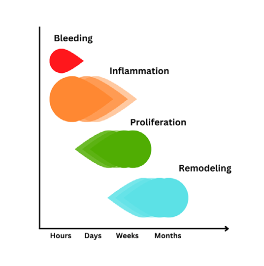
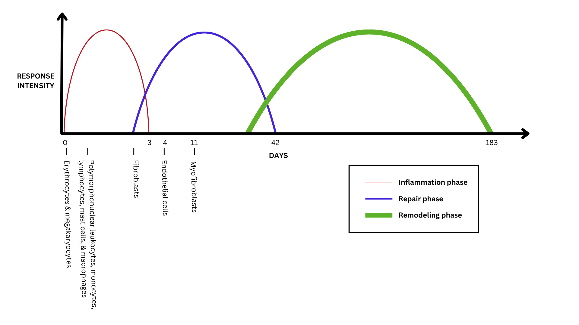
Practitioners must thoroughly understand the phases of healing and pain mechanisms. Backed by science, they can gain the athlete’s confidence, impacting compliance and active engagement in their rehab process. The four phases of healing include hemostasis (bleeding), inflammation, repair (proliferation), and remodeling (see figure 1). The healing process is a continuum, and there is considerable overlap between these three phases.
They involve a cascade of events triggered by chemical mediators in the blood, resulting in a process that varies in length depending on the injury severity. Many factors delay or interrupt this process. Intrinsic factors like systemic medical conditions such as insulin-dependent diabetes mellitus (IDDM) and extrinsic factors like insufficient or inappropriate rehabilitation. It is the practitioner’s responsibility to ensure an optimal environment for the healing process to occur. This involves an accurate sports injury assessment and a large component of athlete education.
Practitioners must guide a graduated return to activity by repeatedly reassessing to avoid re-injury. Soft tissue healing involves a complex interaction between the injured tissues, the vascular system, and chemical mediators to promote damage control, revascularization, and the formation of connective tissue. Inflammation is a healthy, normal, and required local response to injury that destroys or inactivates foreign invaders and sets the stage for tissue repair.
- Hemostasis (bleeding)
When an injury occurs, bleeding happens. Damaged blood vessels constrict to slow blood flow, platelets gather to stop the bleeding, and white blood cells move into the tissue to begin the inflammatory process. This phase is known as hemostasis. - Inflammation
There is considerable cellular activity during inflammation. Neutrophils secrete chemicals to kill bacteria, and macrophages engulf and digest foreign particles and necrotic debris and release angiogenic substances to stimulate capillary growth and granulation (see figure 2). - Proliferation (repair)
Fibroblasts proliferate in the wound and secrete glycoproteins and type III collagen. Epidermal cells migrate from the wound edge, and granulation tissue is formed from macrophages, fibroblasts, and new capillaries. - Remodeling
Fibroblasts secrete collagen to strengthen the scar tissue; collagen remodeling occurs to reorganize fibers, cross-linkages between fibers form, and the scar tissue contracts, increasing tissue integrity.
“Practitioners need to restore the tissue’s biomechanical properties and promote the formation of functional scars.”
Clinical Implications
Chemical and cellular activity during the inflammation seals off the wound while phagocytes move into the inflamed area to destroy bacteria and clear debris from dead and dying cells. Blood supply is re-established due to the dilation of microcirculation vessels and increased permeability of the local capillaries to proteins, and there is visible edema.
Initially, there is vasoconstriction due to the presence of nor-epinephrine and serotonin, but about an hour post-injury, vasodilation allows leucocytes, bradykinins, prostaglandins, and histamines into the area via increased cell permeability, and edema accumulates in cells while fibrin seals off the area.
Late in inflammation, macrophages rid the area of debris, and there is fibroblastic repair of cells and initiation of vascular regeneration. Clinical features include pain at rest and during activity, the latent onset of symptoms, and stiffness, especially following inactivity.
On examination, there is edema, redness, increased local temperature, and resistance through the range, which is limited by a boggy end feel or pain. Symptoms increase with contraction (in the case of muscle strain), stretch, and palpation.
The treatment goals include improving fluid exchange and decreasing edema to control the amount of fibrin produced and limit scar tissue formation. Modalities include POLICE (protect, optimal loading, ice, compression, elevation), active physiological movement to initiate shortening and broadening of the tissue, gentle accessory mobilization, and physiological pump exercises in a shortened position(4).
Wound protection is essential due to the vulnerability of the fragile fibrin network, and it is important to take care not to disrupt the phagocytic activity and formation of new capillaries and collagen tissue with vigorous mobilization.
Table 1: Phases of Healing
| Phase | Duration | Clinical Features | Treatment Goals | Precautions |
| Inflammatory Phase | Hours to 3-5 days | - Edema, redness, warmth. - Pain at rest/activity. - Stiffness after inactivity. |
- Reduce inflammation and edema. - Protect the fragile fibrin network. |
Avoid excessive mobilization to protect capillaries and collagen. |
| Proliferation Phase | 3-6 weeks | - Reduced resting symptoms and stiffness. - Palpable decrease in edema. - Pain limits range of motion. |
- Stimulate circulation and fluid exchange. - Prevent unorganized collagen. - Avoid muscle atrophy. |
Prevent maturation of disorganized collagen. |
| Remodeling Phase | 2-4 weeks to 6-12 months | - Minimal or no resting pain. - Pain with activity or specific positions. - Firm tissue with limited mobility. |
- Restore biomechanical properties. - Promote functional scar formation. - Strength, endurance, and coordination. |
Monitor for re-injury risks due to immature scar tissue. |
Tissue tensile strength increases significantly during the repair phase. Initially, type III collagen fibers have only regained about 15% of their normal tensile strength. Cellular and vascular activities involve cells from the wound margins reproducing and migrating across the wound. This is followed by wound contraction, where the edges of the wound are drawn together. The wound environment stimulates fibroblasts, leading to an increase in collagen production.
The repair phase includes a reduction in resting symptoms, and athletes experience less stiffness. On examination, there is a palpable decrease in edema, normalized temperature, decreased resistance through the range, and a firmer end feel, with a range of motion limited by pain.
The treatment goals in the repair phase include stimulating circulation and fluid exchange to decrease edema and promote healing along functional lines and with sufficient scar length. This means preventing the maturation of unorganized collagen and preserving the soft tissue’s ability to broaden, shorten, and move in relation to surrounding tissues. Deconditioning occurs when athletes are immobilized, and practitioners must ensure they prevent muscle atrophy.
Remodeling can last from 2-4 weeks up to 6-12 months, which gives practitioners and athletes a whole year to influence the scar positively. During this time, collagen synthesis and lysis result in the collagen fibers’ final aggregation, orientation, and arrangement through intra- and extra-molecular cross-linkages. This is when there is also the formation of adhesions, wound contracture, and a return to normal function, but the athlete is still vulnerable to re-injury.
“The healing process is a continuum, and there is considerable overlap…”
Clinical features include minimal to no resting pain and decreased or no latency. Pain is activity or position-dependent, and loss of function is greater than stiffness. On examination, there is very little edema, normal tissue temperature, and firm new tissue on palpation. There may be decreased tenderness on contraction, stretch, and palpation. There should be no through-range resistance, and a firm end feel.
Practitioners need to restore the tissue’s biomechanical properties and promote the formation of functional scars. Treatment options may include more intense accessory movements in a lengthened position utilizing greater force, duration, and amplitude. Additionally, increasing the range of physiological movement, enhancing training to include strength, endurance, and coordination, and incorporating stretching are essential components of effective rehabilitation. Collagen may take 6-12 months to recover full tensile strength.
Conclusion
The injured athlete will experience acute pain. Practitioners should have an in-depth knowledge of this pain mechanism and tissue healing physiology. This knowledge is vital to successfully returning the athlete to sport.
References
1. IJSPT. 2024;19(6):758-767
2. Physical Therapy in Sport 68 (2024) 7–21
3. South African Acute Pain Guidelines Official publication of The South African Anaesthesiologists (SASA)
4. Br J Sports Med January 2020 Vol 54 No 2
Newsletter Sign Up
Subscriber Testimonials
Newsletter Sign Up
Coaches Testimonials
Be at the leading edge of sports injury management
Our international team of qualified experts (see above) spend hours poring over scores of technical journals and medical papers that even the most interested professionals don't have time to read.
For 17 years, we've helped hard-working physiotherapists and sports professionals like you, overwhelmed by the vast amount of new research, bring science to their treatment. Sports Injury Bulletin is the ideal resource for practitioners too busy to cull through all the monthly journals to find meaningful and applicable studies.
*includes 3 coaching manuals
Get Inspired
All the latest techniques and approaches
Sports Injury Bulletin brings together a worldwide panel of experts – including physiotherapists, doctors, researchers and sports scientists. Together we deliver everything you need to help your clients avoid – or recover as quickly as possible from – injuries.
We strip away the scientific jargon and deliver you easy-to-follow training exercises, nutrition tips, psychological strategies and recovery programmes and exercises in plain English.
