You are viewing 1 of your 1 free articles
Sternoclavicular joint dysfunction: a rare diagnosis
Injuries of the glenohumeral joint (GHJ) and the acromioclavicular joint (ACJ) are commonly diagnosed, while injury to the sternoclavicular joint (SCJ) is frequently overlooked during assessments(1). A key reason is that while pain may present locally at the SCJ, it may also occur distally thus replicating referral patterns to that of other joints in close proximity(2). When disorders of the SCJ do occur, they are typically treated conservatively. This is due to the complexity of surgery in this area, with its associated surrounding vital structures(1).
An overview of SCJ injury
An SCJ injury can be described as an anterior/posterior instability of varying severity (i.e. sprain, dislocation or subluxation) and with a varying mechanism of injury (i.e. either via impact or another form of loading)(1). Osteoarthritis (OA) is the most common cause of SCJ pain and swelling. Swelling may be asymptomatic; over 50% of patients over 60 years of age have presented with moderate to severe joint degeneration in cadaveric studies(1). High-risk patients with OA onset are those with a previous dislocation who have chronic instability, post-menopausal women, and manual laborers(1).SCJ injury grading
SCJ injuries can be graded as follows(3):- Mild: Joint is stable and ligament integrity is maintained
- Moderate: Joint subluxation with partial ligament disruption
- Severe: Joint is unstable with complete ligament disruption
Ironically, a minor SCJ sprain is more likely to be diagnosed than a dislocation(1) . Compared to dislocations of the ACJ and GHJ, SCJ dislocations are rarely diagnosed, comprising of just 3% of all diagnosed shoulder girdle dislocations(3). Sternoclavicular joint pain may also be related to degenerative or inflammatory factors (4).
While rarely diagnosed, SCJ dislocations are more frequently experienced in young males due to high-energy collisions, which have a greater potential to cause pain and deformity(4). In a review of 150 SCJ dislocations, 40% were attributed to road traffic accidents (RTA) with 21% related to sports-related injuries. The remaining 39% were related to industrial incidents and falls(5). Anterior dislocations are more common than posteriorly related cases with 98% of cases presenting anteriorly(5).
Though unlikely to occur, 25% of posterior dislocations compromise function of the mediastinal structures, including major blood vessels supplying the upper limb, trachea, esophagus and the phrenic nerve innervating the diaphragm(1). Occlusion to any of these structures could compromise life and referral to an emergency department is therefore essential.
Anatomy and biomechanics
The SCJ is the only synovial joint connecting the upper limb to the axial skeleton (central part of the skeleton – ie head, spine and thorax), and can articulate through three different planes of movement (1).The medial aspect of the clavicle articulates with the supero-medium aspect of the manubrium forming a saddle joint (1). In comparison to many other synovial joints in the body, the bone to bone contact is minimal as less than half of the medial clavicle articulates with the superior angle of the sternum (5).The SCJ responds to movements of the scapula on the chest wall and can elevate through 35 degrees in the frontal plane and 70 degrees in the sagittal plane during abduction(1). The SCJ is stabilized by the anterior and posterior sternoclavicular ligaments, costoclavicular ligament, the interclavicular ligament and the intra-articular disco ligamentous ligament (see figure 1)(4). Researchers have debated whether the costoclavicular ligament or the posterior sternoclavicular ligaments are the primary stabilizers of the SCJ(4).
Figure 1: The anatomy of the SCJ and the associated ligaments
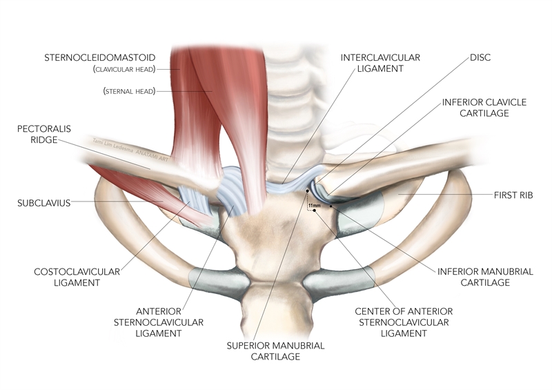
Muscles and ligaments showed left; the right side shows the intra-articular disc and the angle with which the clavicle articulates with the sternum(4).
Examination and assessment
Assess the integrity of the SCJ with the athlete sitting or standing. Observe the clavicle and shoulder complex from all angles(3). Any differences in the contours of the skin surface observed or felt on palpation should raise concerns relating to either fracture of the clavicle or dislocation of the SCJ. Compare both active and passive range of movement of the SCJ and the GHJ across all planes of motion bilaterally. Limitations to range due to significant pain or restriction further point to fracture or dislocation.The brachial plexus lies just posterior to the clavicle. Therefore, any assessment should include a neurological screening, including upper limb tension tests(3). Stiffness, with or without associated pain within the SCJ, can contribute to dysfunction elsewhere in the shoulder girdle. Therefore, the SCJ should be examined if pain presents in the other nearby joints(6).
A patient presenting with an SCJ sprain may describe a situation whereby the shoulder was hit from behind, which forced the shoulder anteriorly(3). An anterior dislocation may occur when a force is applied to the anterolateral shoulder in a posterior direction, with the first rib acting as a fulcrum by which the medial clavicle is forced anteriorly(3,5). An anterior dislocation may also occur as a result of a force applied to the elbow in a medial direction with the shoulder positioned in 90 degrees of abduction(3). In contrast, a posterior dislocation may occur due to a force applied medially, but with the shoulder in a horizontal adducted and flexed position. These types of dislocations can occur in soccer when a player lies under a pile of other players following a goal celebration scenario(3)!
In the event of a suspected dislocation, the patient will most likely undergo an X-ray of the clavicle and the SCJ. Black and colleagues(4)state that some clinicians will request additional CT scans as this reveals the extent of dislocation or fracture, including the posteriorly lying mediastinal structures that may be affected (see figure 2).
Figure 2: CT scan of a posterior SCJ dislocation
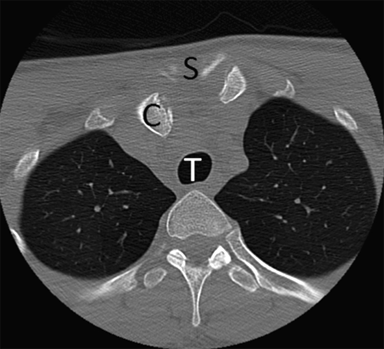
Scan depicts the close proximity of the clavicle in relation to the mediastinal structures such as the trachea. C = Clavicle, S = Sternum and T = Trachea(3). Used with permission bbalcik@hsc.wvu.edu.
Pain referred into the upper trapezius muscle and the shoulder girdle can be derived from numerous structures such as the cervical facet joints, subacromial space and, the ACJ. Researchers from the Concord Hospital in Australia injected hypertonic saline into the SCJ in nine healthy patients to investigate the pain referral patterns produced(2). The results yielded immediate pain presenting within 20 seconds over the SCJ in eight out of nine cases. The majority of patients experienced maximal pain levels within minutes into the anterolateral neck and along the clavicle. One patient experienced pain into the same-sided elbow and jaw too.
Figure 3 shows typical referral patterns in the majority of cases, with the dark red shaded areas producing a greater density of referred pain. Resisted flexion and abduction were also used to determine the effects of pain provocation into the SCJ. Resisted shoulder abduction aggravated the pain in five subjects, had no effect in three cases and alleviated the pain in one case. By contrast, resisted shoulder flexion aggravated the pain in five cases and had no effect in four cases.
Figure 3: Pain referral patterns
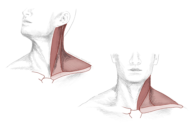 Anterolateral neck (pictured left); Anterior neck and upper shoulder girdle (pictured right)(2).
Treatment and management of SCJ Joint dysfunction
The nature of the injury sustained to the SCJ (i.e. joint sprain or dislocation) will determine whether joint immobilization using a sling is appropriate, or whether more invasive measures are required. Mild sprains should be managed with ice and immobilization in a sling for up to one week and moderate sprains for up to four to six weeks(1). Severe SCJ cases will require closed reduction. This is where the SCJ is relocated under anesthesia, with the patient lying supine, but without the use of invasive open surgical techniques near vital structures(4). To achieve a closed reduction, a towel is positioned between the shoulder blades and the involved arm is abducted to 90 degrees. Traction is applied whilst moving the shoulder into extension towards the floor(3). Pressure is then applied to the medial clavicle in a posterior direction to relocate the SCJ.In the event of SCJ stiffness causing referred pain or dysfunction around the shoulder girdle, passive joint mobilizations to the SCJ can be performed. Maitland has described passive mobilizations in an anteroposterior or longitudinal direction, which allow for effective movement to be achieved into shoulder elevation(6).
In summary
Sternoclavicular joint pain can arise due to the acute onset of a sporting injury, an impact (e.g. caused by a road traffic accident) or a rheumatological disorder. Due to the significant ligamentous stability of this joint, dislocations of the SCJ are rare. However, a posterior dislocation can potentially produce devastating effects due to the vital structures positioned adjacent. Sternoclavicular joint dysfunction is best managed conservatively when possible. Surgical intervention should be considered a last resort due to the delicacy of the surrounding structures. Pain referral patterns of the SCJ dysfunction can mimic other joints in the shoulder; therefore, clinicians should not overlook the SCJ during a clinical assessment.References
- J Bone Joint Surg, June, 2008, 90-B, 6, 685 – 696
- British Soc for Rheu, 2001, 40, 859 – 862
- Prim Care Clin Office Pract, 2013, 40, 911–923
- J Bone and Joint Surg Am, Oct, 2014, 96, 19, e166, 1-10
- Clin Sports Med, 2003, 22, 387–405
- Maitlands peripheral Manipulation, 4thEd, 2005
Newsletter Sign Up
Subscriber Testimonials
Dr. Alexandra Fandetti-Robin, Back & Body Chiropractic
Elspeth Cowell MSCh DpodM SRCh HCPC reg
William Hunter, Nuffield Health
Newsletter Sign Up
Coaches Testimonials
Dr. Alexandra Fandetti-Robin, Back & Body Chiropractic
Elspeth Cowell MSCh DpodM SRCh HCPC reg
William Hunter, Nuffield Health
Be at the leading edge of sports injury management
Our international team of qualified experts (see above) spend hours poring over scores of technical journals and medical papers that even the most interested professionals don't have time to read.
For 17 years, we've helped hard-working physiotherapists and sports professionals like you, overwhelmed by the vast amount of new research, bring science to their treatment. Sports Injury Bulletin is the ideal resource for practitioners too busy to cull through all the monthly journals to find meaningful and applicable studies.
*includes 3 coaching manuals
Get Inspired
All the latest techniques and approaches
Sports Injury Bulletin brings together a worldwide panel of experts – including physiotherapists, doctors, researchers and sports scientists. Together we deliver everything you need to help your clients avoid – or recover as quickly as possible from – injuries.
We strip away the scientific jargon and deliver you easy-to-follow training exercises, nutrition tips, psychological strategies and recovery programmes and exercises in plain English.


