Plantaris tendon: the nuisance bystander?

The plantaris muscle (PM) is a small, thin, and spindle-shaped muscle (1.5 x 7-13 cm in length) located in the posterosuperior aspect of the lower leg(1,2). The PM appears as a vestigial muscle, which is absent in 7-20% of limbs(3,4). Along with the soleus and gastrocnemius, it is part of the muscle group known collectively as the triceps surae.
Anatomy and function
The PM originates on the lateral supracondylar line of the femur, just above and slightly medial to the origin of the lateral head of the gastrocnemius, and lateral to the popliteal vessels and tibial nerve (see figure 1)(1). Cadaver studies reveal the origin of the PM can have many variations, including blending with the oblique popliteal ligament(1,5,6). From its origin, the PM courses downwards in an inferior and medial direction across the popliteal fossa. It has a long thin tendon (mean length varying from 24.7cm to 35cm) that runs between the medial head of the gastrocnemius and soleus muscle in the middle of the leg(1,7).Figure 1: Anatomy of the plantaris

Because the plantaris tendon (PT) is long and sleek, novice students mistake it for a nerve during cadaver dissections; hence it’s moniker as the ‘freshman’s nerve’ or ‘fool’s nerve’(2,8). The tendon continues along the medial aspect of the Achilles tendon (AT) and inserts in up to nine different anatomical variations(9-12). These anomalies include insertion into the AT itself, the fascia of the lower leg, the plantar aponeurosis, or the flexor retinaculum of the ankle. The most common insertion, however, is a wide fan-shaped placement medial to the PT on the calcaneal tuberosity(11,13).
The AT does not have a synovial sheath as other tendons in the body; instead, it has a paratendon, which is a thin, fibrous, and highly vascularized band of tissue. The PT runs within this paratendinous sheath, and this may be a potential site of adhesions between the PT and AT(1,14). The PM is a weak plantar flexor of the ankle and flexor of the knee, and its main role may be proprioceptive - as evidenced by the large numbers of muscle spindles contained within the small body of the muscle(1,2,8,15).
Injury to the plantaris
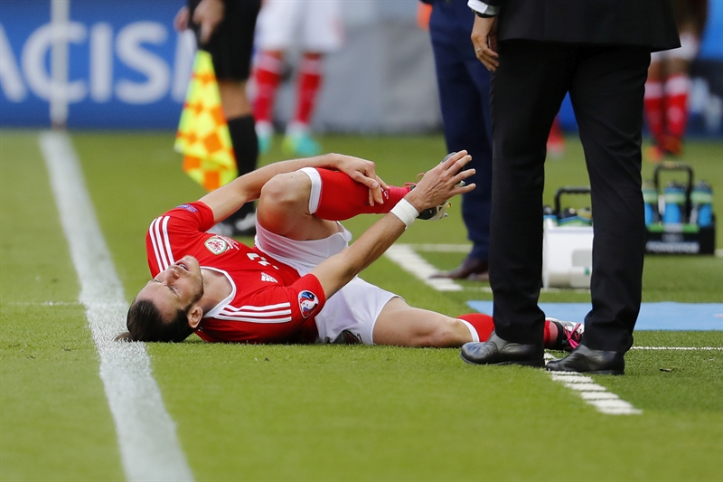
The mechanism of injury to the PM may be similar to that of the gastrocnemius - a sudden eccentric load while moving into ankle dorsiflexion with the knee extended - as occurs in jumping and sprinting (16,17). The athlete may feel like they’ve been hit in the back of the knee or upper calf. However, they may not lose power when running and jumping. A tear in the medial gastrocnemius (sometimes referred to as tennis leg) can occur as well(18). The next day, the calf may present with extreme soreness, and bruising may track down the inside of the calf muscle. Dorsiflexion (both passive and active) and resisted plantar flexion produce pain.A plantaris injury may occur as an isolated injury, in combination with a soleus and gastrocnemius tear, or an ACL injury(5,6). Damage is more common in the proximal muscle belly or the musculotendinous junction but infrequently occurs through the tendon(18,19). Ruptures isolated to the distal tendon are tender to palpation two to three centimeters above the calcaneal insertion on the medial side of the AT and are typically mildly swollen(20).
Relationship to Achilles tendinopathy
In recent years, there has been some interest in the relationship between mid-portion Achilles tendinopathy and the presence of an enlarged PT, which may compress the AT. The reasoning is as follows:- The PT is stiffer and stronger than the AT and thus may create an interface problem due to excessive shear and compression between the two tendons(21).
- After surgical removal of the PT, the tendon structure in the AT gradually improves(22,23).
The symptoms of plantaris-related Achilles pain differ from classic Achilles tendinopathy. Athletes with the plantaris-related variety usually participate in sports that require explosive full-range ankle joint movements and complain of sharp pain on the medial side of the AT during push-off. Furthermore, moving from loaded plantar flexion to dorsiflexion, such as when lowering from a calf raise, may elicit pain(13).
In some symptomatic cases, when the AT was investigated in surgery, the PT was found fused to the AT at the same location as the reported pain(24). This anatomical variation creates an area of compression between the PT and the AT, rather than a natural gliding action between the two. The results of many studies largely support this view:
- A potential compression zone exists between the AT and PT, due to an invagination of the PT into the AT(1).
- Ultrasound and Doppler-guided imaging confirm the co-location and enlargement of the PT and subsequent pathology in 80% of patients who had mid-portion Achilles tendinopathy(25).
- In most patients who complained of mid-portion, medial-sided Achilles tendinopathy, the PT was found close to (and in some cases invaginated into) the mid portion of the AT (also confirmed prior to surgery with radiology)(23).
- A small surgical cohort of elite and recreational athletes who complained of medial sided Achilles pain in the absence of notable signs of AT pathology (such as thickened tendons, imaging findings on ultrasound), found that surgical removal of the PT improved post-surgical symptoms(13).
Imaging
MRI and ultrasonography are both suitable for investigating muscle or tendon tear, or the involvement of the PT in AT pathology. An MRI evaluation may show(19):- A high signal from T2 weighted images in and around the muscle belly in the popliteal fossa or at the myotendinous junction.
- Complete ruptures as a retracted mass between the popliteus tendon and the lateral gastrocnemius, or as an intermuscular hematoma between the soleus muscle and medial head of gastrocnemius.
Ultrasound reveals a hyper-echoic structure with an internal fibrillar appearance(26). Using ultrasound or MRI in the detection of PT-related AT pathology is also useful. In this instance, signs of a classic tendinopathy in the AT appear very close to the area where the PT pushes against the AT.
Injury management
*Tears to the muscle, myotendinous junction, and distal tendonRecovery from isolated tears of the PM or myotendinous junction follows the same time course as more common tears of the gastrocnemius. An injury may initially require a period of protected weight bearing if walking and ambulation are difficult. However, because the muscle is small and bears only a weak function, rehabilitation usually progresses quickly due to the lack of limitations. Furthermore, tears of the distal tendon do not seem to be that debilitating. A case study in Switzerland described an isolated tendon rupture in an athlete who returned to professional football in four weeks with minor training restrictions early in the post-injury period(20).
*Association with Achilles tendinopathy
Management of PT-related AT pain is not well described in the literature. Due to the bulk of PT-related papers being surgically biased, the reader might assume that the only way to manage this problem is with surgery. If a true compression zone exists between the PT and AT - and this is what causes the AT pathology - then removal of the compression zone through surgery may appear to be the only solution. However, a PT-related AT pathology may also respond to the classic loading protocols of AT tendinopathy. The only caveat may be to avoid full dorsiflexion positions, which create large amounts of compressive force.
In summary
Plantaris muscle and tendon injury is an uncommon occurrence in elite athletes. It may present similarly to a gastrocnemius tear or it may be implicated in chronic cases of Achilles tendinopathy. Management of these problems follows similar guidelines for muscle injury and tendinopathy management.References
- The Bone & Joint Journal. 2016. 98-B(13), 12-19
- Moore KL, Dalley AF, eds. Clinically Oriented Anatomy. 5th ed. Philadelphia: Lippincott Williams & Wilkins, 2006; 648–649
- J Hand Surg [Am]. 1991; 16:708–711
- Sports 2019, 7, 124; doi:10.3390/sports7050124 1-10
- Radiology 2002; 224:112–119
- Radiology 1995; 195:201–203
- AJR 1999; 172:185–189
- J Can Chiropr Assoc 2007;51:158–165
- Res. Int. 2018, 2018
- Radiol. Anat. 2017, 39, 69–75
- Surg Gynecol Obstet. 1946. 83:107–116
- Anat. 2011, 218, 336–341
- BMJ Open Sport Exerc Med 2018;4:e000462. doi:10.1136/bmjsem-2018-000462
- J Foot Ankle Surg. 2011. 40, 132–136
- Scand J Med Sci Sports 2000; 10:312–320
- Sports Med 1999; 27(3):193–204
- Clin in Sports Med 1988; 7(2):435–437
- Radiol (2011) 40:891–895
- Skeletal Radiology · December 2010 DOI: 10.1007/s00256-010-1076-0
- The Journal of Foot & Ankle Surgery 2018. 57 (2018) 995–996
- J Foot Ankle Surg. 2011;17:252–5
- BMJ Open Sport Exerc Med. 2015;1: e000005
- BMC Musculoskeletal Disorders 2016; 17:97. 1-6
- Foot Ankle Clin 2006; 11, 429–438
- J. Sports Med. 2011, 45, 1023–1025
- Br J Plast Surg. 1990;43:689–91
You need to be logged in to continue reading.
Please register for limited access or take a 30-day risk-free trial of Sports Injury Bulletin to experience the full benefits of a subscription. TAKE A RISK-FREE TRIAL
Newsletter Sign Up
Subscriber Testimonials
Dr. Alexandra Fandetti-Robin, Back & Body Chiropractic
Elspeth Cowell MSCh DpodM SRCh HCPC reg
William Hunter, Nuffield Health
Newsletter Sign Up
Coaches Testimonials
Dr. Alexandra Fandetti-Robin, Back & Body Chiropractic
Elspeth Cowell MSCh DpodM SRCh HCPC reg
William Hunter, Nuffield Health
Be at the leading edge of sports injury management
Our international team of qualified experts (see above) spend hours poring over scores of technical journals and medical papers that even the most interested professionals don't have time to read.
For 17 years, we've helped hard-working physiotherapists and sports professionals like you, overwhelmed by the vast amount of new research, bring science to their treatment. Sports Injury Bulletin is the ideal resource for practitioners too busy to cull through all the monthly journals to find meaningful and applicable studies.
*includes 3 coaching manuals
Get Inspired
All the latest techniques and approaches
Sports Injury Bulletin brings together a worldwide panel of experts – including physiotherapists, doctors, researchers and sports scientists. Together we deliver everything you need to help your clients avoid – or recover as quickly as possible from – injuries.
We strip away the scientific jargon and deliver you easy-to-follow training exercises, nutrition tips, psychological strategies and recovery programmes and exercises in plain English.


