You are viewing 1 of your 1 free articles
Pectoralis Minor
Chris Mallac outlines the anatomy and biomechanics of pectoralis minor, how tightness can create injuries to the shoulder, and also effective stretching and loosening procedures for this muscle.
Chicago Bulls forward Tyson Chandler (L) is fouled by Phoenix Suns forward Amare Stoudemire as Chandler attempts to take a shot, 2004.
The pectoralis minor (PMi) is a muscle found on the anterior chest wall that directly affects movement of the scapula, an important consideration for proper scapulohumeral movement (see figure 1). It has been suggested that tightness in the PMi can adversely affect scapula function, specifically in limiting upward rotation, external rotation, and posterior tilting, leading to shoulder injuries such as impingement syndrome, rotator cuff pathologies, internal impingement, glenohumeral instability, and adhesive capsulitis(1-9).

Figure 1: Pectoralis minor anatomy
Anatomy and biomechanics
PMi is a broad triangular shaped muscle that arises from the second to fifth ribs and their costal cartilages via aponeurotic slips that are continuous with the facia covering the intercostal muscles(10). In an old but thorough cadaver study, Anson found that 42% of these slips are attached from the second to the fifth ribs(10). But this origin may be reduced to three or even to two ribs between the second to fifth ribs.
Overall 91.5 % of cadavers studied showed that the PMi arose from all or some of the ribs between the second and the fifth. Cases where the origin extended beyond the territory of the second to the fifth ribs made up but 8.5 % of the total number.
From here the fibres course upwards and laterally and converge to insert onto the anteromedial and superior surface of the coracoid process. The PMi is found deep to the more superficial pectoralis major muscle, while the axillary blood vessels and brachial plexus lie posterior to the muscle (see figure 1).
In a further study, it was found that in some cadaver specimens, the PMi tendon has an ectopic insertion, that is, somewhere away from the corocoid process(11). These ectopic insertions were described as being of three varieties:
- Type 1 – occurs when the entire PMi tendon passes over the superior margin of the coracoid process to insert distally on multiple sites such as the supraspinatus tendon, the coracoacromial ligament, one of the tubercles of the humeral head, or the labrum.
- Type 2 – this variation is seen when some portion of the tendon has normal insertion into the coracoid process and another portion has anomalous insertion.
- Type 3 – occurs when the muscle itself (as opposed to the tendon) inserts anomalously without attaching to the coracoid process.
This study also suggested that patients with an ectopic insertion had significantly thicker fibrotic scar tissue in the rotator interval adjacent to the coracoid process. These scar changes around the coracoid process including the rotator interval may have a different pathological or physiological mechanism that causes adhesive capsulitis. The overlying tendon running on top of the coracoid process could cause friction against the coracoid process or structures in the rotator interval during motion.
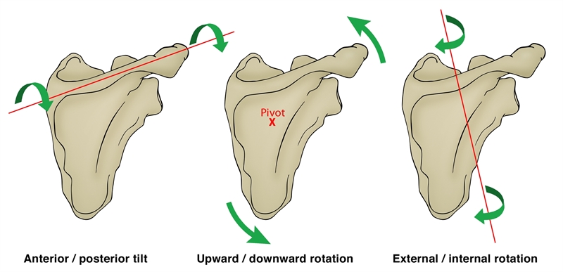
Figure 2: Scapular kinematics
PMi functions
The functions of the PMi (see figure 2) are to:
- Protract the scapula (along with the serratus anterior).
- Downwardly rotate the scapula (along with rhomboids and the levator scapulae).
- Anterior tilt the scapula (along with rhomboid and levator scapulae).
- Depress the scapula (along with the inferior trapezius).
- Internally rotate the scapula.
- Assist the inspiration muscles during breathing (due to its attachment onto the rib cage).
Sporting/ training activities that involve these motions in the scapula (racquet sports, swimming, weight lifting etc) can theoretically result in overuse of this muscle, especially in the presence of training errors, poor technique or a rapid increase in training load, frequency and/ or duration. Furthermore, the muscle can become habitually shortened due to postural positions such as sitting at a computer or constant reaching with one hand. These may all lead to tightness in the muscle and this will prevent full scapular motion during overhead movements as it may limit upward rotation and posterior tilt of the scapula, movements that are necessary for clearance of the acromion process away from the humeral head during arm elevation(3).
Pectoralis minor length measurement
A number of tests have been proposed to measure length in the PMi. These include:
Test 1 - Stand erect with the test arm resting at the side. Use a tape measure to measure the linear distance between the origin and insertion of the muscle. The origin can be defined as the inferior aspect of the 4th rib, which is one finger width lateral to the sternum, just lateral to the sternocostal junction. The insertion is defined as the medial-inferior aspect of the coracoid process. This method of measuring PMi length has previously been proven to be valid when compared with measurements made using an electromagnetic tracking system. The complications of this test are that it measures from the inferior part of the coracoid, whereas the PMi inserts onto the superior part. Furthermore, the PMi has a broad attachment across ribs two to five. Therefore measuring from the 4th rib may be insufficient and not be an accurate reflection of the muscle’s origin. Finally, it can be difficult to properly palpate the PMi due to the bulky pectoralis major.
Test 2 - This test involves measuring the linear distance from a treatment table to the posterior aspect of the acromion process. Lie supine on a standard treatment table and adopt a naturally relaxed posture. Place the arms by the sides, and the elbows flexed and resting against the lateral wall of the abdomen. The hands rest gently on the abdomen, which places the glenohumeral joint in slight internal rotation – about 6cm is a normal expected range for the acromion process. However, larger posterior deltoids will ‘push’ the acromian process further away from the table; therefore, a better measure may be to simply compare sides and ascertain if the symptomatic shoulder has a greater distance implicating PMi as a tight muscle in the pathology.
Investigators have also studied the more effective stretches for the PMi in producing increases in length. A ‘unilateral’ self-stretch produces the greatest changes in length. This stretch is performed in a standing position with the target shoulder abducted to 90° and the elbow flexed to 90°. With the hand’s volar surface of the target arm placed against a vertical door frame or other flat rigid structure, the subject then rotates their trunk away from the target shoulder. This stretching maneuver places the humerus in an abducted and externally rotated position while stretching the shoulder further into horizontal abduction and sequentially retracting the scapula. A stretch to the PMi would require movement of the muscle’s insertion in a posterior direction in conjunction with a scapular retraction that is performed at or above 30° of flexion or elevation in the scapular plane (scaption), thereby lengthening the muscle. Arm elevation angles that are 30° or greater would require the scapula to upwardly rotate and further posteriorly tilt to provide additional lengthening of the otherwise shortened muscle.
Injuries and tightness in PMi
Due to its attachment to the coracoid process, a shortening of PMi will lead to anterior tilting and downward rotation of the scapula and prevent full upward rotation, elevation, and posterior tilting of the scapula, which is required for full shoulder elevation. A number of clinical syndromes are associated with a shortening of PMi(12). These include; thoracic outlet syndrome, scapular winging and tilting syndrome, scapular abduction syndrome, scapular depression syndrome, and scapular downward rotation syndrome. Moreover, athletes may experience insertional tendinopathy of PMi caused by bench pressing and called this ‘Bench Presser’s Shoulder’(13). 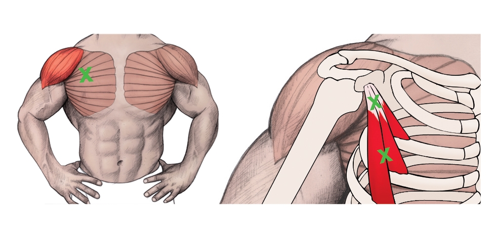
Figure 3: Trigger Points in the PMi
PMi trigger point referral
Trigger points in the PMi can also create pain in and around the shoulder (see figure 3)(14). The pain referral pattern of PMi is mostly over the anterior deltoid area and spills over into the subclavicular and pectoral regions. The pain may extend down the medial aspect of the arm, forearm, and into the ulnar distribution of the hand and the third, fourth, and fifth digits. Furthermore, PMi trigger points may create pain similar to angina(15).
Moreover, tightness in PMi can also be a causative factor in thoracic outlet syndrome(16). Thoracic outlet syndrome is the result of compression or irritation of neurovascular bundles as they pass from the lower cervical spine into the arm via the axilla. If the PMi muscle is involved, the patient may present with chest pain, along with pain and paraesthesia in the arm. The symptoms were reproduced on both digital pressure over the PMi muscle and on provocative testing for thoracic outlet syndrome. Treatment should focus on the PMi muscle.
Stretching exercises for PMi
Self Stretch
a. Place the 90° flexed arm against a wall with the hand closed into a fist.
b. The shoulder angle is approximately 60-70°. This is less than 90° as this degree of shoulder abduction involves the pectoralis major too much, and therefore this may become the limiting factor. The 60-70° position may be sufficient to gain enough upward scapular rotation and posterior tilt to adequately impart a stretch onto PMi. The humerus can now be horizontally extended to create scapula retraction to fully stretch the PMi.
c. Slowly rotate the body away from the stretch arm to increase the retraction.
d. Hold for 30-45 seconds and repeat for three repetitions.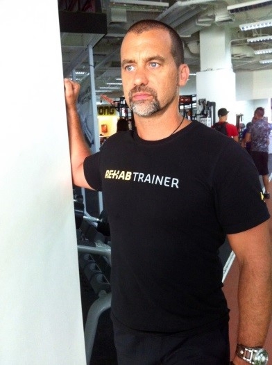
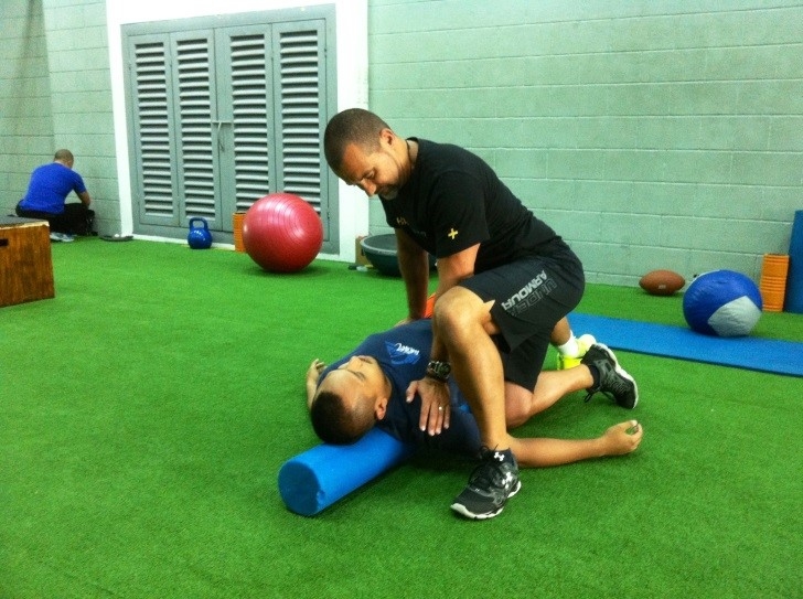
Partner assisted stretch
a. Have the subject lie length-wise on a foam roller. This ensures the scapulae are free to retract and not fixed by the floor.
b. Rest the hand in supination/external rotation on the floor at 30-45° abduction.
c. The trainer/therapist can impart a force onto the coracoid process to create a retraction, posterior tilt, upward rotation, and elevation movement. These four movements are opposite to what the PMi does when it contracts.
d. Notice the angle of the stretching arm – it is angled to push the corocoid not only into retraction, but also into upward rotation and elevation.
e. To gain extra stretch, the patient breathes in deeply, and then the stretch is applied during expiration.
f. Hold the stretch for 5 seconds and release, allowing the patient to breathe in again and repeat. This can be repeated for 10-15 breaths.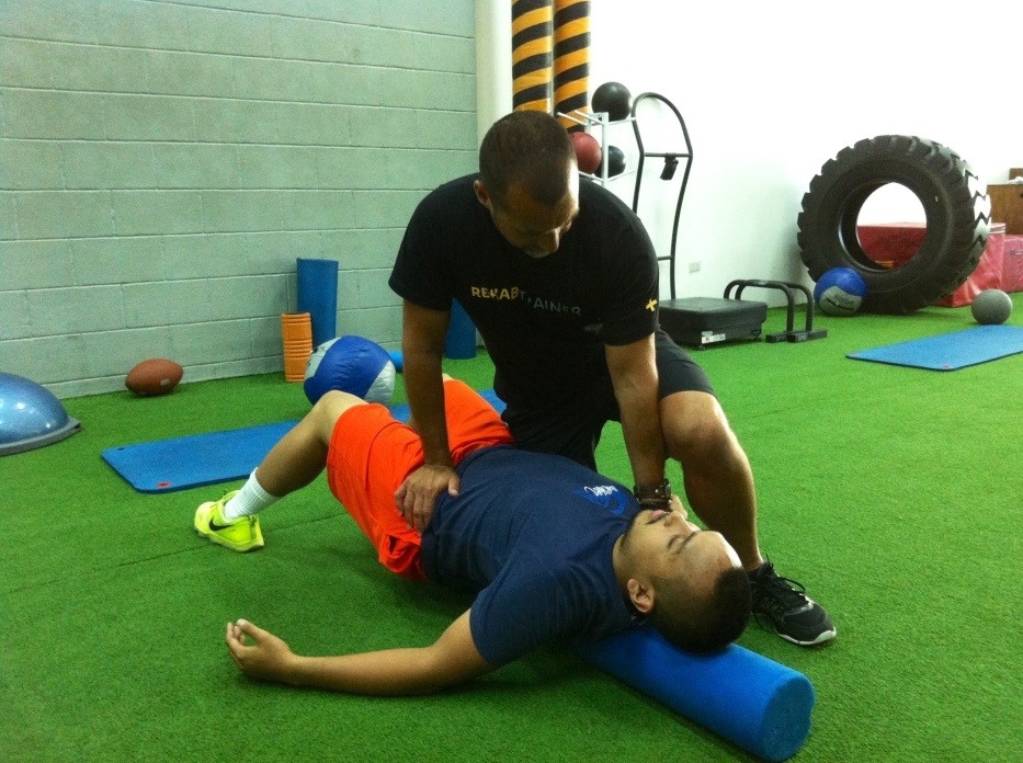
Trigger point release
a. Using any manner of trigger point release devices (in this example, two taped tennis balls are being used), place it against the upper chest below the clavicle and slightly medial to the anterior deltoid. This ‘triangle’ of tissue is where the coracoid process and PMi can be found.
b. Place the arm on the releasing side into external rotation. This encourages scapula retraction.
c. Gently lean into the release device, searching for any areas of tightness and tension. Hold for 20-30 seconds, remove, and move the device elsewhere and repeat.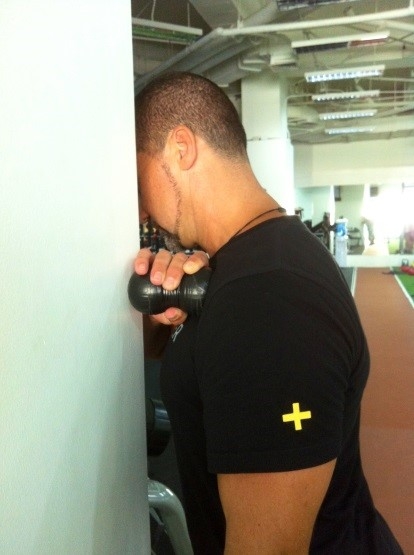
Summary
The PMi has been identified as a muscle that is implicated in scapula dysfunction and subsequent shoulder pathologies. Tightness in the muscle can restrict full scapula upward rotation, posterior tilt, and elevation – movements that are required to fully allow clearance of the acromian process away from the humeral head in full elevation positions. Stretching exercises for the PMI has been offered to lengthen and reduce tone through this problematic muscle.
References
- Phys Ther. 2000; 80(3):276-291
- Sports Medicine and Arthroscopy Review. 2012; 20(1):39
- J Orthop Sports Phys Ther. 2005; 35(4):227- 238
- Phys Ther. 2009; 89(4):333-341
- J Orthop Res 2001; 19(6):1192-1198
- J Shoulder Elbow Surg. 2005;14(1 Suppl S):58S-64S
- J Orthop Sports Phys Ther. 2006; 36(7):485- 494
- Am J Sports Med. 2007; 35(8):1361-1370
- J Biomech. 2008; 41(2):326-332
- J.Anat. 1938; 72(4); 629-630
- Korean J Radiol. 2014;15(6):764-770
- Sahrmann S: Diagnosis and treatment of movement impairment syndromes. London: Mosby; 2002
- Br J Sports Med. 2007 Aug;41(8):e11
- Simons DG, Travell JG, Simons LS. Travell & Simons’ myofascial pain and dysfunction. The trigger point manual volume 1. Upper Half of Body. 2nd ed. Baltimore, MD: Williams & Wilkins; 1999
- J Chiropractic Med 2011; 10, 173–178
- J Can Chiropr Assoc. 2012 Dec;56(4):311-5
Newsletter Sign Up
Subscriber Testimonials
Dr. Alexandra Fandetti-Robin, Back & Body Chiropractic
Elspeth Cowell MSCh DpodM SRCh HCPC reg
William Hunter, Nuffield Health
Newsletter Sign Up
Coaches Testimonials
Dr. Alexandra Fandetti-Robin, Back & Body Chiropractic
Elspeth Cowell MSCh DpodM SRCh HCPC reg
William Hunter, Nuffield Health
Be at the leading edge of sports injury management
Our international team of qualified experts (see above) spend hours poring over scores of technical journals and medical papers that even the most interested professionals don't have time to read.
For 17 years, we've helped hard-working physiotherapists and sports professionals like you, overwhelmed by the vast amount of new research, bring science to their treatment. Sports Injury Bulletin is the ideal resource for practitioners too busy to cull through all the monthly journals to find meaningful and applicable studies.
*includes 3 coaching manuals
Get Inspired
All the latest techniques and approaches
Sports Injury Bulletin brings together a worldwide panel of experts – including physiotherapists, doctors, researchers and sports scientists. Together we deliver everything you need to help your clients avoid – or recover as quickly as possible from – injuries.
We strip away the scientific jargon and deliver you easy-to-follow training exercises, nutrition tips, psychological strategies and recovery programmes and exercises in plain English.


