You are viewing 1 of your 1 free articles
Navigating Sports Medicine’s ‘Bermuda Triangle’ – Part I
Groin pain remains a challenging sports injury to manage. The complex regional anatomy provides practitioners with the unenviable task of diagnosing and managing athletes with groin pain. However, a universal terminology and taxonomy would assist practitioners in diagnosing and managing these athletes through the return to play process. In part I, Candice MacMillan discusses the international consensus on groin pain terminology, assessment, and diagnosis.
Introduction
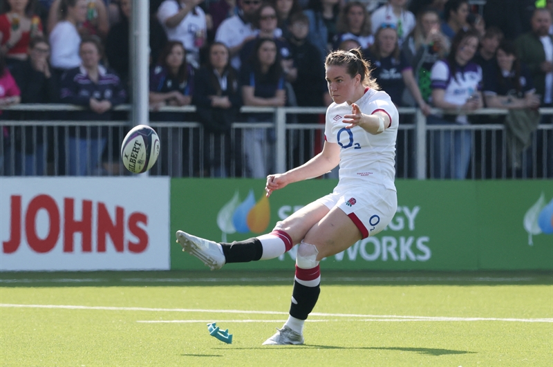
Athletic groin pain is often triggered or exacerbated by sports that require rapid acceleration and deceleration, change of direction, and forceful hip movements such as kicking(1,2). While symptoms could be mild, groin pain could develop into a career-altering disability if misdiagnosed and treated inappropriately.
Traditionally, clinicians consider the groin the ‘Bermuda Triangle’ of sports medicine(2,3). The complexity of the underlying pathology, intricate regional anatomy, and limited consensus among researchers results in controversies among practitioners regarding the assessment, diagnosis, and treatment of groin pain. In addition, referred pain from related visceral pathology and other joints such as the hip, sacroiliac joint (SIJ), and lumbar spine further complicates the assessment and treatment plan(2,4). However, in recent years, researchers have attempted to provide uniform guidelines for the clinical examination, diagnostic imaging, and treatment to optimize athletes’ eventual return to play (RTP)(1,2,5,6). The Doha agreement meeting on terminology and definitions in groin pain in athletes attempts to standardize the groin-related pain terminology and provide definitions for clinical practice.
Assessment
Subjective assessment
Practitioners need to identify red flags during the subjective evaluation. Red flags include a history of fever, trauma, unexplained weight loss, night pain, painful urination, or prolonged corticosteroid use(1,2). Red flags coupled with a history of prostate or reproductive organ cancer and abdominal and pelvic organ disorders are the reason for referral to relevant specialists for further investigation.
Furthermore, information regarding the athlete’s age, sport, and level of participation are relevant. For example, adolescent athletes participating in sports that require high load activities such as kicking and sprinting are at risk of avulsion fractures more than adults due to the open epiphyseal growth plates(2).
Physical evaluation
The physical evaluation of the groin requires a thorough clinical examination which includes palpation, pain provocation, joint ROM assessment, muscular strength, and sport-specific function(2,7). The Doha agreement meeting provides practitioners with a framework to assess and diagnoses groin pain according to the location of pain on palpation or provocation (see figure 1)(8,9).
| 5 | Examiner can’t break participant’s initial isometric hold. |
| 4 | Examiner can break participants’ initial isometric hold, but maximum resistance by the participant after the initial break. |
| 3 | Examiner can break the initial isometric hold and move the participant through the range but against moderate resistance from the participant. |
| 2 | Examiner can break the initial isometric hold and move the participant easily through the range with minimal resistance from the participant. |
| 1 | Examiner can break the initial isometric hold with minimal effort and no resistance offered by the participant through range. |
_________________________________________________________________
Functional testing
It is common for athletes with groin pain to continue training and playing with mild groin pain. Most athletes typically seek medical assistance when the pain impacts their sports participation or performance. Training and playing with groin pain can result in movement-compensation strategies, which practitioners can identify during the physical evaluation.
The objective assessment of impairments and function can include bent-knee fall-out for testing hip range, single-leg stance, single-leg squat, Star Excursion Balance Test for testing balance and mobility, the timed 10-m cutting sprint (5-m sprint with 75° cut and 5-m sprint finish) or T-agility test (see figure 3)(2). However, further evidence is needed to create standard clinical performance-related tests for athletes with groin pain. Therefore, practitioners should select various tests that best simulate the sports demands of the injured athlete.
Figure 3: Timed 10-m cutting test

The athlete sprints 5 meters with a 75° cut and then sprints 5 meters to finish. Compare the time to complete a sprint between the affected and unaffected cutting leg.
Lumbar spine and SIJ screening tests
Practitioners can rule out discogenic or radiculopathy pathology of the lumbar spine without peripheral or central symptoms, normal lumbar ROM, and a negative slump and straight leg raise test. In addition, practitioners can use the thigh thrust test to rule out SIJ pathology(2).
Differential diagnosis
Whether acute or chronic, groin pain can be, and is usually, multifactorial. For example, strains of muscles attaching to the pelvic girdle, bony injury to the pelvic girdle, the inguinal canal or hip, and lumbar spine pathology can be associated with groin pain (see table 1)(2,4,14).
Table 1: Differential diagnosis in athletes with groin pain(1,2,3,15)
| Bone injury | Musculotendinous | Visceral, Neural & Other | |
|---|---|---|---|
| Acute | Trauma-related fractures: | Adductor longus (kicking/COD) | Sciatic nerve entrapment |
| Femoral head or neck | Rectus Femoris (kicking/sprinting) | Ilioinguinal nerve entrapment | |
| Acetabulum | Iliacus (COD) | Referral from L4-L5 (discogenic or radiculopathy) | |
| Public ramus | Psoas (COD) | Reproductive organ or prostate cancer | |
| Iliacus ischium | Rectus abdominis | Inguinal hernia | |
| Pubic symphysis | Abdominal hernia | ||
| Avulsion fracture of ASIS or SIS | Femorolacetabular impingement (FAI) syndrome | ||
| Slipped capital femoral epiphysis | Referral from SIJ | ||
| Acetabular labral tear | |||
| Chronic | Femoral head avascular necrosis | Tendinopathy: | |
| Osteoarthritis (older athletes) | Adductor longus | ||
| Hip and pelvic stress fracture | Rectus Femoris | ||
| Pelvic apophysis injury (adolescents) | Iliopsoas | ||
| Pubic apophysitis (adolescents “ mid 20’s) |
*ASIS= Anterior superior iliac spine; SIS = Superior Iliac Spine; COD = Change of Direction
Imaging
There is currently limited evidence to suggest that imaging improves the diagnosis or prognosis of groin pain(2,7). If practitioners consider the multifactorial nature of longstanding groin pain, imaging findings can rarely determine the cause of pain(7). While imaging often shows morphological load-related changes, the challenge is to understand which of these are related to the specific pathology(1,7). Excessive imaging may result in athletes unnecessarily focusing on normal morphological changes, making them fearful of movement and impeding effective treatment(2,7). Currently, imaging of players with longstanding groin pain is most helpful to rule out severe potential pathology.
For acute groin pain, imaging might be more helpful. MRI is slightly more sensitive than ultrasound for acute groin injuries and indicated for adductor-, pubic- and hip-related groin pain(1,2,7). Ultrasonography is appropriate for investigating inguinal- and iliopsoas-related groin pain. Although the best modality to show the apophyses is computed tomography, clinicians should be cautious with youth athletes due to high doses of radiation(2). X-rays are helpful for the diagnosis of FAI syndrome, fractures, and the identification of degenerative changes in the hip(1,2).
Conclusion
The Doha classification of groin pain provides a valuable guideline to practitioners regarding possible causes of groin pain. Unfortunately, high-quality conclusive research regarding assessment and specific treatment modalities is still scarce. However, there is consensus on the terminology and definitions of groin-related pain. The agreement provides Sports and Exercise Medicine professionals with a common language that should improve the multi-disciplinary treatment of athletes with groin pain.
Reference list
- Br J Sports Med. 2016;50(19):1181-1186.
- J Orthop Sports Phys Ther. 2018;48(4):239-249.
- Acta Clin Croat. 2014;53(4):471-478.
- Orthop J Sports Med. 2018;6(5):232596711877167.
- J Sci Med Sport. 2018;21(10):999-1003.
- Br J Sports Med. 2015;49(22):1447-1451.
- Aspetar Sports Med J. 2021;10.
- Br J Sports Med. 2015;49(12):768-774.
- J Sci Med Sport. 2022;25(1):3-8.
- Br J Sports Med 2015;49:803–9.
- Br J Sports Med 2015;49:810
- Sports Med. 2017;47(10):2011-2026.
- Phys Ther Sport. 2021;52:272-279.
- Co-Kinet J. 2018;(78):7-7.
- Muscle Ligaments Tendons J. 2019;05(03):214.
Further reading
Newsletter Sign Up
Subscriber Testimonials
Dr. Alexandra Fandetti-Robin, Back & Body Chiropractic
Elspeth Cowell MSCh DpodM SRCh HCPC reg
William Hunter, Nuffield Health
Newsletter Sign Up
Coaches Testimonials
Dr. Alexandra Fandetti-Robin, Back & Body Chiropractic
Elspeth Cowell MSCh DpodM SRCh HCPC reg
William Hunter, Nuffield Health
Be at the leading edge of sports injury management
Our international team of qualified experts (see above) spend hours poring over scores of technical journals and medical papers that even the most interested professionals don't have time to read.
For 17 years, we've helped hard-working physiotherapists and sports professionals like you, overwhelmed by the vast amount of new research, bring science to their treatment. Sports Injury Bulletin is the ideal resource for practitioners too busy to cull through all the monthly journals to find meaningful and applicable studies.
*includes 3 coaching manuals
Get Inspired
All the latest techniques and approaches
Sports Injury Bulletin brings together a worldwide panel of experts – including physiotherapists, doctors, researchers and sports scientists. Together we deliver everything you need to help your clients avoid – or recover as quickly as possible from – injuries.
We strip away the scientific jargon and deliver you easy-to-follow training exercises, nutrition tips, psychological strategies and recovery programmes and exercises in plain English.
