You are viewing 1 of your 1 free articles
Masterclass: AC joint reconstruction - Part I
In part 1 of this two-part feature, Chris Mallac outlines the relevant anatomy and biomechanics of the accromio-clavicular joint, how they are injured, how to clinically assess injury and the relevant radiological requirements in determining the extent of injury.

Injuries to the acromioclavicular joint (ACJ) are reasonably common injury in the athlete and recreational pursuits such as cycling. It is a common complaint in contact sport athletes such as rugby, AFL, NFL and combat sports such as judo and MMA. In fact, Headey et al. (2005) show that in elite-level rugby union in the United Kingdom, ACJ injuries account for 32% of shoulder injuries, being the most frequent structure to be injured playing Rugby Union.
How these injuries are managed remains controversial. Low-grade ACJ sprains are managed conservatively; however, the more serious ACJ separations can be managed surgically or non-surgically. Bosworth, in 1941 offered the first surgical intervention into serious ACJ dislocations. Today numerous surgical techniques are available to repair a dislocated ACJ.
Relevant anatomy and biomechanics
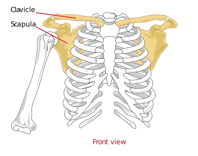
The joint between the clavicle and the acromion (AC joint) and the sternum and the clavicle (SC joint) are the only connections the appendicular skeleton (arms) has to the axial skeleton (trunk). Therefore the acromioclavicular joint (ACJ) may be subject to high forces due to this unique anatomical arrangement.
The ACJ is a diarthrodial joint with four planes of movement, anterio-posterior (forwards and backwards) and superoinferior (up and down). It is also able to spin on its axis during shoulder movements. The joint is surrounded by a capsule and has an intra-articular synovium making this a synovial joint. Both the clavicular and acromial bones are covered with cartilage (hyaline in late teens and early twenties that matures into fibrocartilage in the 20s).
The ACJ also has an intervening meniscoid disc between the two bones. The exact function of the meniscoid disc is poorly understood and the disc is not as well defined as the meniscoid disc that is housed within the sternoclavicular joint (SCJ). This may explain why the disc degenerates quickly with age. By the age of 40, it may be non-existent (Peterssen 1983). Due to the small nature of this disc and the high compressive forces encountered at the joint due to muscle contraction of the powerful shoulder muscles such as the pectoralis major and latissimus dorsi, this disc is thought to be prone to early breakdown along with the distal end of the clavicle. Both the disc and distal clavicle are prone to compressive failure, evidenced by the high rate of osteolysis in the distal clavicle, especially in athletes who impose huge forces on the joint such as weightlifters (Richards 1993).
The ACJ is supported by four AC ligaments – superior, inferior, anterior and posterior. These ACJ ligaments prevent excessive antero-posterior movement. Furthermore, ligaments join the coracoid process to the clavicle (the coracoacromial ligaments – CC ligaments) and these are the trapezoid and conoid ligaments. These ligaments provide supero-inferior support (up and down) as well as anterior translation support.
When the acromion separates from the clavicle (fall onto the shoulder), the ACJ ligaments are the first ligaments to stretch and withstand force (in particular, the superior ACJ ligaments), followed then by the conoid ligament and trapezoid ligament. Therefore, injuries to only the AC ligaments may still be considered a stable injury whereas injury to the conoid/ trapezoid ligaments will result in there being disruption of the ACJ as well as part of the CC ligament complex.
In 1986, Fukuda et al performed a study on cadaver specimens that investigated the contribution these ligaments play in ACJ stability. What they found can be summarised below:
- The AC ligament acts as a primary restraint to posterior clavicle displacement and posterior axial rotation;
- The conoid ligament constrains anterior and superior rotation as well as anterior and superior displacement of the clavicle.
Furthermore, Rockwood (1998) states that the superior AC fibers blend with the fibers of the upper trapezius and deltoid which attach to the superior aspect of the clavicle and acromion, and as a result, he argues these muscles may be important in providing active support to the ACJ.
Injuries to the ACJ
Injuries to the ACJ are far more common in men in their early twenties, highlighting behavior as a pre-determining factor in ACJ injury. Young men are more commonly involved in sports and pastimes that will potentially lead to a traumatic ACJ injury. Sports such as football, rugby, motocross riding, mountain bike riding and combat fighting (judo, MMA, jujitsu) are sports where there exists a potential for traumatic ACJ injury.
The most common mechanisms of injury are direct falls onto the shoulder whereby the acromion is driven into the ground and the clavicle separates from the acromial attachment. This can happen in contact situations in football sports or falls off a bike whereby the rider lands on the shoulder.
When the acromion contacts the ground, a downward displacement of the clavicle is primarily resisted through an interlocking of the sternoclavicular ligaments (Bearn 1967). The clavicle remains in its normal anatomic position, and the scapula and shoulder girdle are driven inferiorly. The result, then, of a downward force being applied to the superior aspect of the acromion is either give-way of the AC and CC ligaments or clavicle fracture. There may be an additional anteroposterior direction to the force. If the downward force continues, tears of the deltoid and trapezius muscle attachments occur from the clavicle, as well as ruptures of the CC ligament. With severe force, the skin overlying the AC joint can also be disrupted. In the rare type VI injury to the AC joint, a different mechanism of injury is responsible. A severe direct force onto the superior surface of the distal clavicle, along with abduction of the arm and retraction of the scapula, has been described. The clavicle is driven inferiorly, where it lodges beneath either the acromion or the coracoid.
Indirectly, a fall onto an outstretched arm may force the humeral head to push up against the acromion, thus resulting in an ACJ separation. This is known as an indirect injury. These types of injuries only affect the AC ligaments and the CC ligaments remain intact.

Figure 1: Anatomy of the acromioclavicular joint
Clinical evaluation
Usually, the mechanism of injury is quite obvious – acute ACJ pain following a direct fall onto the point of the shoulder. Pain will be felt directly over the ACJ and gross swelling may be evident, as well as an obvious step deformity, highlighting a probable high-grade ACJ injury.
Most objective measures, such as range of movement (particularly horizontal adduction), will be pain-limited, and the ability to generate isometric rotator cuff strength will also be pain-limited. In low-grade injuries, often the athlete is able to achieve 80%+ of movement; however, more serious grade 2 injuries onwards will be pain-limited.
Table 1: Grading system for ACJ injury
| Type | Features |
| I | The AC ligaments are sprained, but the joint is intact. No palpable placement of the joint itself. Minimal to moderate tenderness and swelling over the AC joint. Patients have only minimal pain with movement of the arm. Radiographically there may be mild soft tissue swelling, but there is no widening, separation, or deformity at the AC joint. |
| II | AC ligaments are torn, but the CC ligaments are intact. Type II injuries are characterized by moderate to severe pain at the AC joint. The distal end of the clavicle may be palpated to be slightly superior to the acromion and shoulder motion produces more pain at the AC joint. The distal clavicle is also found to be unstable in the horizontal plane if grasped and moved anterior to posterior. A bilateral Zanca view may demonstrate that the distal clavicle is slightly elevated, but the CC interspace is the same in both the injured and uninjured shoulders. |
| III | Patients with type III injuries present with the upper extremity in a supported, adducted and elevated position to help relieve pain. In type III injuries both the AC and CC ligaments are torn, but the deltoid and trapezial fascia are intact. The distal clavicle may be prominent enough to tent the skin and is unstable in both the vertical and horizontal planes. These patients have a severe amount of pain with tenderness to palpation at the AC joint. Any movement of the arm, especially abduction, creates pain and discomfort, especially for the first one to three weeks. Both plain and bilateral Zanca x-rays reveal that the distal clavicle is 100% displaced superiorly in relation to the acromion. In actuality, the position of the clavicle is not altered by the injury. The weight of the upper extremity causes the acromion to displace inferiorly in relation to the horizontal plane of the lateral clavicle. In obvious cases of dislocation, the clavicle is displaced superiorly from the acromion and the CC interspace will be greater in the injured shoulder. |
| IV | Type IV injuries are characterized by complete dislocation with posterior displacement of the distal clavicle into or through the fascia of the trapezius. Physical examination of these patients reveals a greater amount of pain as compared with patients with type III injuries,and the pain is located more posteriorly. Examination of the seated patient from above will reveal that the distal clavicle is displaced posteriorly when compared with the uninjured shoulder. It is possible for the distal clavicle to become ˜button-holed in the trapezius and tent the skin posteriorly. With a type IV injury, it is also important to examine the SC joint for a concomitant anterior dislocation. The posteriorly displaced clavicle is best appreciated on an axillary view ofthe shoulder. |
| V | Type V injuries represent a greater degree of soft tissue damage, with the delto-trapezial fascia being stripped off the acromion and the clavicle. These injuries present as a more severe type III injury with more pain and a greater amount of displacement at the AC joint. The distal end of the clavicle appears to be grossly displaced superiorly toward the neck. The scapula is translated anteriorly and inferiorly as it migrates around the thorax. On the bilateral Zanca view, there is a 100-300% increase in the CC interspace. Patients with a type V injury may have pain in the neck or trapezius due to the disruption of the delto-trapezial fascia. |
| VI | Type VI injuries are inferior AC joint dislocations into a subacromial or subcoracoid position. Type VI injuries are usually seen in high-energy polytrauma patients. The mechanism of injury is extreme hyperabduction and external rotation of the arm combined with retraction of the scapula. Associated injuries include clavicle and upper rib fractures and upper root brachial plexus injuries. It is not uncommon for these patients to have transient paraesthesias that subside after reduction. |
Radiography
A number of exposures are required to properly visualize the ACJ and the associated structures. This can be best described under the following points:
- Reduce the X-ray penetration to a third to a half of that commonly used for a shoulder X-ray.
- Zanca view is the best for visualizing the ACJ. This is done by tilting the X-ray beam 10-15° cephalad and imaging both ACJ on the same cassette to compare the coracoclavicular distance (CC distance).
- The axillary view is required for Type IV injuries where the distal clavicle has been posteriorly displaced.
- A Stryker notch view is the best view for visualizing coracoid process fractures.
- Stress views using 10-pound weights attached to the wrist (rather than holding in the hands) are used to exacerbate the ACJ separation.
Tossy et al. (1968) and Rockwood (1998) provide the most comprehensive grading system for ACJ injury, and these are typed from Type I-VI (see table 1).
Management of AC injuries
Typically, Type I ACJ separations are minor injuries and are generally treated conservatively. This may involve a short period of immobilization with a sling for five to seven days to reduce the stress on the AC joint however, this is not common across all sports. Regular ice is applied for the first 48 to 72 hours. As with most acute soft tissue injuries, non-steroidal anti-inflammatory drugs (NSAIDs) are usually avoided for the first 3-4 days due the potential for delayed healing of collagen structures (Warden 2005). Immediate isometric and gentle range-of-motion exercises are encouraged. A more structured strengthening program is initiated as soon as the patient’s symptoms begin to resolve.
Gross power movements (gym-based) can be slowly progressed, usually in the following order:
- Horizontal pulling movements (seated row, prone fly, bent over row);
- Vertical pulling – elbows in front of the shoulder (close grip pull-downs, hammer grip chin-ups, straight arm pull-downs);
- Horizontal pushing (bench press, dumbbell bench press);
- Vertical pushing (shoulder press movements);
- Diagonal PNF patterns.
Type II injuries usually involve more extensive soft tissue trauma. Treatment for type II injuries is essentially the same as for type I injuries, although the time frame is prolonged due to the greater trauma sustained. A period of immobilization will almost always be required. A sling is generally used for 1 to 2 weeks. The patient is informed that a mild cosmetic deformity may be present once the injury is healed.
The time frame for a return to sport is varied depending on the severity of the initial injury, the type of sport played and associated previous shoulder injuries. Simple type one injuries may be back to sport within 7-10 days; more severe type II injuries may take up to eight weeks to return to full function. The focus in part two of this article will be on the rehabilitation following surgical fixation of the ACJ in type III-VI injuries.
Conclusion
ACJ injuries are common injuries in the contact sport athlete and in recreational pursuits whereby the sufferer falls onto the point of the shoulder. The ACJ is surrounded by a collection of AC and CC ligaments that may fail sequentially, leading to varying types of ACJ injury.
The more benign type I and II injuries can usually be managed conservatively. The type III injuries form the ‘difficult to decide’ category for conservative versus surgery, whereas type IV, V, and VI all need surgical intervention.
Part II of this masterclass outlines in detail the surgical options available to reconstruct a Type IV-VI injury and the typical post-operative rehabilitation.
Further reading
Newsletter Sign Up
Subscriber Testimonials
Dr. Alexandra Fandetti-Robin, Back & Body Chiropractic
Elspeth Cowell MSCh DpodM SRCh HCPC reg
William Hunter, Nuffield Health
Newsletter Sign Up
Coaches Testimonials
Dr. Alexandra Fandetti-Robin, Back & Body Chiropractic
Elspeth Cowell MSCh DpodM SRCh HCPC reg
William Hunter, Nuffield Health
Be at the leading edge of sports injury management
Our international team of qualified experts (see above) spend hours poring over scores of technical journals and medical papers that even the most interested professionals don't have time to read.
For 17 years, we've helped hard-working physiotherapists and sports professionals like you, overwhelmed by the vast amount of new research, bring science to their treatment. Sports Injury Bulletin is the ideal resource for practitioners too busy to cull through all the monthly journals to find meaningful and applicable studies.
*includes 3 coaching manuals
Get Inspired
All the latest techniques and approaches
Sports Injury Bulletin brings together a worldwide panel of experts – including physiotherapists, doctors, researchers and sports scientists. Together we deliver everything you need to help your clients avoid – or recover as quickly as possible from – injuries.
We strip away the scientific jargon and deliver you easy-to-follow training exercises, nutrition tips, psychological strategies and recovery programmes and exercises in plain English.


