You are viewing 1 of your 1 free articles
Hip, hip, hooray! Athletes don’t have to call it a day
Developmental hip dysplasia has the potential to derail the physical development of athletes at all levels. However, with early identification and accurate treatment, athletes can return to competitive sports and remain physically active for years to come. Candice MacMillan discusses the diagnosis and management of DHD and provides guidelines on getting athletes back to sports.
Developmental hip dysplasia (DHD) is a disorder associated with anomalies in the articular and periarticular anatomy of the hip. The associated structural abnormalities result in hip instability, capsular laxity, and abnormal growth of the acetabulum(1,2). Subsequently, the biomechanics of the hip are affected. The multifactorial etiology includes genetic, environmental, and mechanical risk factors(2).
Developmental hip dysplasia is common in sports such as ballet, gymnastics, figure skating, competitive cheerleading, and wrestling(3,4). Athletes who participate in these sports use their hips in a supraphysiologic, mechanically complex manner. From a performance perspective, increased joint range of motion and flexibility associated with DHD is a favorable attribute until joint deterioration occurs(3). Repetitive movement in these extreme ranges of motion results in compensatory osseous and muscular pathology(1,3,5). Whether innate or acquired, these athletes have some hypermobility predisposing them to hip impingement or instability(4,5). Athletes with inherent generalized hyperlaxity are at increased risk of injury and may require longer recovery times to return to performance (RTP). If left untreated, symptomatic DHD leads to early-onset osteoarthritis. Both nonoperative and operative treatments (arthroscopic and open) have an essential role in returning athletes to their preoperative performance levels(1,3,6).
Anatomy and pathology
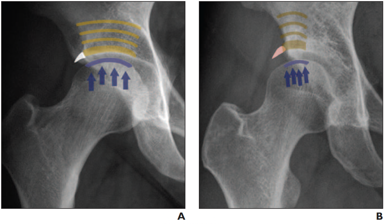
The hip is a synovial, ball-and-socket joint formed by the femoral head that fits into the acetabulum. The development of the acetabulum and femoral head is closely interrelated, and interference during infancy or early childhood affects the congruency of these structures leading to DHD(1,4). More specifically, DHD is a disorder in which the abnormal development of the hip results in a shallow acetabulum with limited anterior and lateral cover of the femoral head (see figure 1)(2).
A: Anteroposterior X-ray of a normal acetabular roof. Note that weight bearing forces (blue arrows) are applied over the entire surface. B: Dysplastic shallow acetabular roof with weight bearing forces distributed over a smaller area.
Prolonged maligned contact also leads to chronic changes like hypertrophy of the capsule and ligament teres and forming a thickened acetabular edge(2,3). These changes further affect joint congruency and prevent the appropriate relocation of the femoral head. As a result, DHD includes unstable hips, malformed acetabula, subluxation, and dislocation (see figure 2).
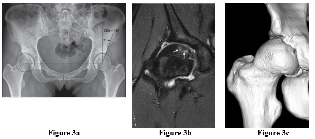
Figure 3a: A-P X-ray indicating the LCEA used to diagnose the severity of hip dysplasia
Figure 3b: Coronal MRI image reveals an enlarged lateral labrum with substantial deterioration (black arrow) and subchondral changes in the femoral head (white arrows)
Figure 3c: 3D CT images further illustrate the combined pattern of cam and femoral coverage.
The diagnosis of DHD-related bony dysmorphism is not always clear. In addition, hybrid conditions may exist with varying degrees of dysplasia, chondolabral pathology, and femoroacetabular impingement(3,5). Therefore, determining the underlying etiology and most appropriate intervention can be challenging.
Diagnosis
A complete medical history, physical examination, and X-ray or MRI evaluation are required to diagnose symptomatic hip dysplasia and other associated pathology. Some athletes may have a treatment history for hip problems as an infant or child. However, symptoms may recur in adulthood if there is some remaining deformity. Common symptoms of hip dysplasia include:
- Intermittent pain in the groin, hip, buttock, and thigh. The pain usually subsides with rest.
- A sensation of catching, popping, clicking, or giving way with activity.
- Worsening pain with sitting, walking, or running
- Limping
- Increased difficulty with strenuous activities
On X-ray examination, clinicians assess several angles to measure femoral coverage. For example, a lateral center-edge angle (LCEA) of less than 20° is defined as severe dysplasia, 20-25° degrees, borderline dysplasia, and >25°is normal(7). Additional CT or MRI scans are required for a thorough diagnosis of associated pathology not visible on X-ray (see figure 3).
A: X-ray of an 18-year-old male patient with acetabular dysplasia (shallow socket) of both hips before PAO.
B: X-ray of both hips in the same patient after correction of the shallow sockets with PAO surgery. Note the improved coverage of the femoral head.
Image sourced from: www.ortho.wustl.edu/content/Patient-Care/3205/Services/Hip-Knee/Adult-Reconstruction-and-Hip-Preservation-Overview/Hip-Dysplasia.aspx
Treatment
Depending on the severity of DHD-related symptoms and dysfunction, associated pathology, the disorder progression, and the patient profile, rehabilitation with or without surgery is a treatment option. The primary treatment goal is to obtain a concentrically positioned femoral head into the acetabulum, structuring or stimulating the latter to adapt normally.
Surgical intervention
Clinicians may consider joint preservation surgery if hip dysplasia is diagnosed early (before osteoarthritis). However, if hip dysplasia is diagnosed later in the disease process and osteoarthritis is present, joint preservation surgery may not be an option, and clinicians may consider a total hip replacement (THR). Notably, DHD is the main cause of THR in younger athletes. A periacetabular osteotomy (PAO) is the preferred surgical intervention for symptomatic DHD(6,8). A PAO is an open surgery that re-orientates the dysplastic acetabulum and may provide a lifelong solution for symptomatic dysplasia (see figure 4)(6,8). Most athletic patients undergoing PAO are female and show postoperative improvements in function and return to athletic play(6).
Considering the associated structural pathology, it is essential to differentiate the principal etiology of joint pain and subsequent dysfunction in athletes. Clinicians can use arthroscopy to correct borderline dysplasia and femoral acetabular impingement (FAI) while correcting severe dysplasia requires an open procedure(3,8). Successful outcomes are achieved with arthroscopic correction of FAI, and athletes’ probability of returning to sports is high. However, inappropriate arthroscopy in the presence of dysplasia can lead to direct surgery-related instability and potentially unfavorable consequences(3).
Rehabilitation
Post-hip arthroscopy rehabilitation can start as early as one day after surgery. Depending on the extent and type of the procedure, rehabilitation programs should be tailored to meet the individual’s needs. Patients use crutches to limit weight bearing and a brace for stability to limit flexion to 90° and extension to 0°(9,10). A daily stationary bicycle is recommended for eight weeks post-operatively, after which clinicians can progressively include impact-type exercises such as jogging.
Researchers at the Aarhus University Hospital in Denmark conducted an eight-week supervised progressive resistance training program to increase muscle strength and induce muscle hypertrophy for patients with dysplasia awaiting POA surgery(11). The program included 3-4 sets of 8-12 repetitions of exercises, such as the leg press, hamstring curl, knee extension (all performed double-legged), hip flexion (single-legged), and lunges(11). The study results demonstrate that supervised progressive resistance training is viable in improving pain, functional, muscle strength in patients with hip dysplasia(11). However, this study included a non-athletic population.
Inadequate rehabilitation after PAO may lead to poor outcomes, including prolonged impairments in hip strength(12). Therefore, developing standard PAO rehabilitation programs is vital for achieving optimal outcomes. A panel of experts in North America developed rehabilitation guidelines for use following a PAO (see table 1)(12).
Table 1: Rehabilitation guidelines post PAO surgery(12)
|
Weight bearing precautions and discharge of crutches |
· 25%, foot-flat weight bearing for 6-8 weeks. · Concomitant procedures, such as hip arthroscopy or microfracture, may prolong these recommendations. · Discharge crutches after 6-8 weeks of protected weight bearing. · Observe gait deviations prior to crutch discharge. |
|
Range of motion precautions |
· Hip flexion and external rotation ROM should be protected for 4-6 weeks followed by progressive, pain-free restoration of ROM. · End range stretching can be initiated between 8-12 weeks as tolerated with full ROM achieved by 12-16 weeks post-operatively. |
|
Protection of the hip flexor complex |
· Progressive loading of the hip flexor complex should be done cautiously, with isometrics and short-lever active assistive hip flexion exercises beginning between 4-8 weeks as indicated by pain. · Long-lever active hip flexion should be avoided for 8-12 weeks following a PAO. |
|
Lumbopelvic and posterior-lateral hip strengthening |
· Start in the early postoperative phase in non-weight bearing and progress to double and single leg weight bearing exercises as tolerated. |
|
Lumbopelvic and lower extremity neuromuscular control |
· Start in non-weight bearing in the early postoperative phase with progression to weight bearing exercises at 6-8 weeks or immediately after the patient is cleared for weight bearing. · In weight bearing, exercises should consist of double and single-leg exercises challenging frontal plane and femoral internal rotation control. |
|
Return to sports criteria |
· The upright stationary bike can be initiated between 6-8 weeks, and the elliptical trainer between 10-12 weeks to facilitate early-stage cardiovascular endurance and passive range of motion of the hip. · Utilizing a combination of strength, endurance, and functional performance measures during return-to-play testing, including but not limited to hip abductor-to-adductor strength ratios, the Y-Balance test, and various hop tests. |
Conclusion
Hip dysplasia is usually a progressive condition that worsens with time. Hip pain, dysfunction, and activity limitations worsen as the condition progresses. Therefore, early diagnosis and consideration of different treatment options are important. Athletes generally respond well to arthroscopic and PAO surgery combined with rehabilitation and can return to high-level competition post-operatively.
References
- Children. 2022;9(2):247.
- Developmental Dysplasia Of The Hip. StatPearls Publishing; 2022.
- J Hip Preserv Surg. 2017;4(4):332-336. doi:10.1093/jhps/hnx028
- Sports Health Multidiscip Approach. 2015;7(4):346-358.
- BMJ Open Sport Exerc Med. 2021;7(4):e001169.
- Am J Sports Med. 2016;44(6):1573-1581.
- Am J Roentgenol. 2013;200(5):1077-1088.
- Am J Sports Med. 2014;42(8):1791-1795.
- Am J Sports Med. 2021;49(9):2447-2456.
- EFORT Open Rev. 2019;4(9):548-556.
- J Rehabil Med. 2018;50(8):751-758.
- Int J Sports Phys Ther. 2022;17(6). doi:10.26603/001c.38043
Newsletter Sign Up
Subscriber Testimonials
Dr. Alexandra Fandetti-Robin, Back & Body Chiropractic
Elspeth Cowell MSCh DpodM SRCh HCPC reg
William Hunter, Nuffield Health
Newsletter Sign Up
Coaches Testimonials
Dr. Alexandra Fandetti-Robin, Back & Body Chiropractic
Elspeth Cowell MSCh DpodM SRCh HCPC reg
William Hunter, Nuffield Health
Be at the leading edge of sports injury management
Our international team of qualified experts (see above) spend hours poring over scores of technical journals and medical papers that even the most interested professionals don't have time to read.
For 17 years, we've helped hard-working physiotherapists and sports professionals like you, overwhelmed by the vast amount of new research, bring science to their treatment. Sports Injury Bulletin is the ideal resource for practitioners too busy to cull through all the monthly journals to find meaningful and applicable studies.
*includes 3 coaching manuals
Get Inspired
All the latest techniques and approaches
Sports Injury Bulletin brings together a worldwide panel of experts – including physiotherapists, doctors, researchers and sports scientists. Together we deliver everything you need to help your clients avoid – or recover as quickly as possible from – injuries.
We strip away the scientific jargon and deliver you easy-to-follow training exercises, nutrition tips, psychological strategies and recovery programmes and exercises in plain English.


