Hamstring or not? nerve entrapment and posterior thigh pain
Identifying the difference between focal or referred posterior thigh pain is critical in developing the appropriate management strategy. Jo Brown explores posterior thigh pain and helps clinicians to determine when a hamstring injury may be a canary in the coal mine for nerve entrapment.
Hamstring injuries account for approximately 12% of all reported sporting injuries. In sprinters, the incidence is as high as 29%, with a recurrence rate of 32%(1). Hamstring injuries present in one of two ways, complete or partial tear during high-speed running or under acute stretch, which commonly occurs in dancers. In addition, the hamstrings often get blamed for pain that is not all their fault. Therefore, accurate diagnosis is imperative in the appropriate management of these cases. The athlete’s history provides many clues to a non-hamstring origin of pain. The broad spectrum of etiological factors for posterior thigh pain includes acute trauma, iatrogenic inflammatory conditions, infectious diseases, vascular abnormalities, and gynaecologic and other space-occupying lesions. Furthermore, clinicians must rule out lumbosacral pathology and other intrapelvic lesions in athletes with sciatic nerve symptoms(2).
A comprehensive subjective examination should guide differential diagnosis between an actual hamstring injury and alternate causes of posterior thigh pain (see table 1)(3).
Table 1: Hamstring strain vs. referred pain
|
Hamstring strain |
Referred pain |
|
Sudden or severe pain |
Sudden or gradual tightness/pain |
|
Disabling- mobility affected |
Athletes complain of a ‘cramp’ or ‘twinge’ |
|
Decreased strength/stretch |
Athletes may be able to walk or jog pain-free |
|
Marked focal tenderness |
Often non-specific tenderness |
|
Possible local bruising/hematoma |
Slump often positive |
|
Possible lumbar and SIJ signs |
May have lumbar signs, and gluteal palpation may reproduce pain |
Clinicians must be cautious when assessing athletes when differentiating between a local strain and referred pain. For example, elite runners commonly complain of a ‘cramp’ or ‘twinge’ that clinicians may consider a low-grade strain. However, some diagnostic distinctions include gradual onset, no focal point of tenderness, and unaffected gait or jogging(4). Accurate differential diagnosis and management require an in-depth anatomical understanding of the posterior thigh region.
Nerve entrapment - where?
Sciatica is a colloquially used term to collectively describe the posterior leg pain syndrome caused by the compromised sciatic nerve. However, specific pain patterns exist for different levels of entrapment. Previously known as piriformis syndrome, sciatica is the entrapment of the sciatic nerve as it passes between the piriformis and deep hip rotators. In addition, it is often associated with trauma and limited sitting tolerance(5).
The next level of entrapment is ischiofemoral impingement. This occurs as the nerve gets compressed between the lateral ischial tuberosity and the lesser trochanter at the level of quadratus femoris (and other deep rotators). Athletes may report pain while walking, particularly in mid- to terminal stance(6). As the sciatic nerve clears the pelvis, it can become entrapped by the proximal or distal hamstrings(7). Therefore, clinicians should maintain a high index of suspicion, particularly when the athlete has a history of hamstring injuries. In addition, possible partial avulsion or thickening of the hamstring at any point may entrap the sciatic nerve(8,9). The Straight Leg Raise and Slump tests assess the neurodynamic status of the sciatic nerve.
Historically, clinical symptoms associated with piriformis syndrome were thought to be caused by prolonged and excessive contraction of the piriformis muscle. However, now we have a better understanding of hip kinematics and anatomy. As a result, the clinical evaluation has evolved, and thus the understanding of sciatic nerve mechanics. In addition, other areas within the deep gluteal space have now been identified(10). Although piriformis syndrome is still widely used, these observations led to the introduction of the term deep gluteal syndrome (DGS). It better distinguishes the pathophysiology and clinical symptoms of hip, buttock, and thigh pain and radicular pain caused by non-discogenic and extra pelvic entrapment of the sciatic nerve and its branches.
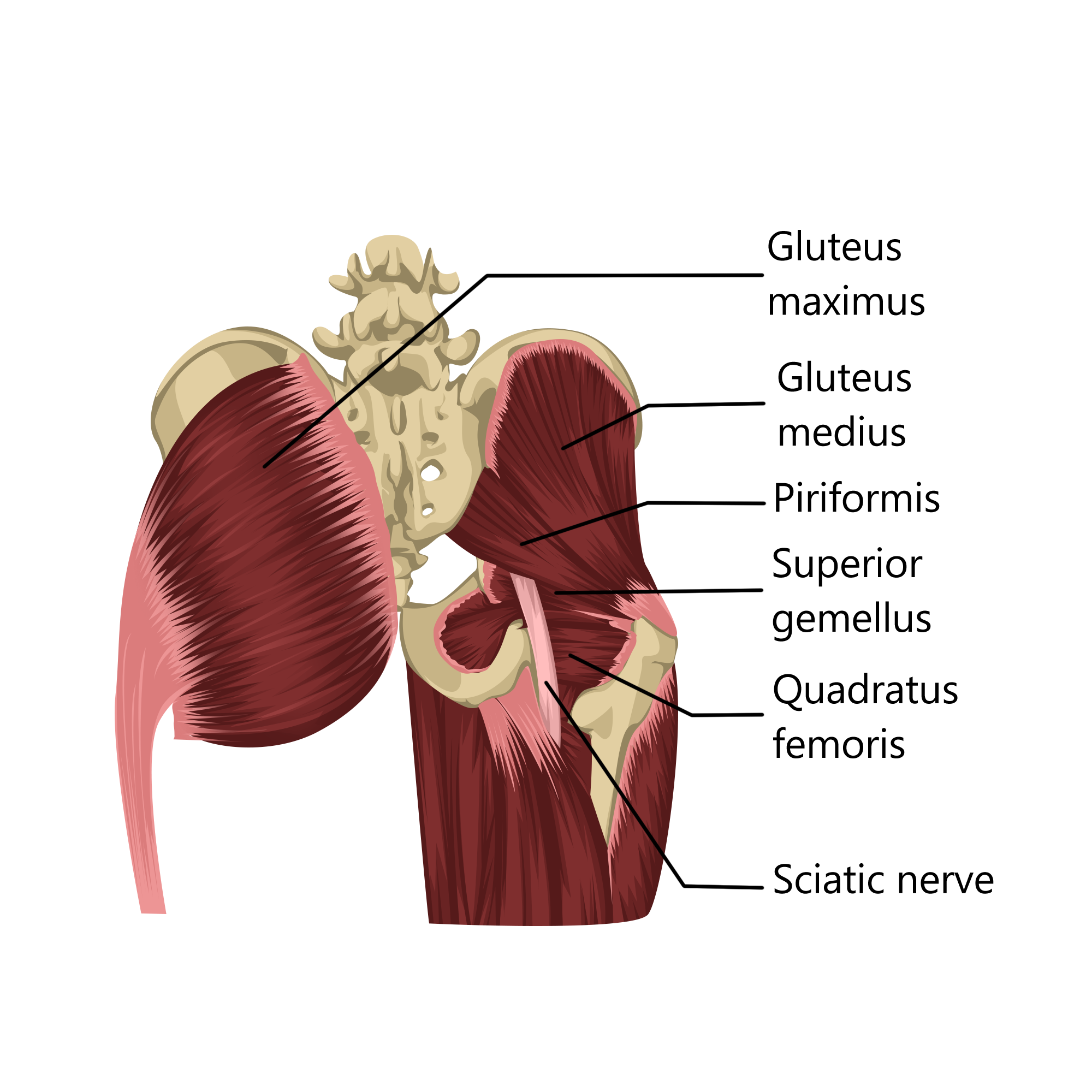
Early descriptions of sciatic nerve irritation were attributed to the anatomic relationship between the piriformis muscle and the sciatic nerve. On the other hand, DGS may be associated with radicular-type pain due to a non-discogenic sciatic nerve entrapment in other structures within the subgluteal space (see figure 1)(11).
The short lateral hip rotators include piriformis, obturator internus, gemelli, and quadriceps femoris. These muscles may be involved in sciatic nerve entrapment. This may be due to an anatomic abnormality or post-traumatic scarring and fibrous bands that entrap or restrict sciatic nerve movement(12).
Both quadratus femoris and ischiofemoral syndrome are due to impingement of the ischium and femur, which results in abnormal contact between the lesser trochanter and the ischium. This is usually secondary to trauma but can be associated with large ranges of hip motion in certain sports. In addition, narrowing the space between the ischial tuberosity and femur causes hip pain and may contribute to sciatic nerve entrapment(13).
In the posterior thigh, the sciatic nerve runs superficial to the adductor magnus but deep to the hamstring muscle complex, which consists of the biceps femoris, semitendinosus, and semimembranosus. In addition, the sciatic nerve lies approximately 1.2 cm lateral to the ischial tuberosity or the outer border of the semimembranosus tendon. Thus, the proximal part of this muscle complex is extremely close to the sciatic nerve.
Proximal hamstring tendon pathologies can cause irritation or entrapment of the sciatic nerve because of the intimate relationship between the nerve and muscle at the level of the ischial tuberosity. Many cases have been associated with repetitive stress on the hamstring tendon and reported in sporting activities that involve running, kicking, or jumping. Furthermore, MRI observations demonstrate sciatic nerve pathology in two-thirds of patients following a hamstring injury and significant impairment in sciatic conductivity(14). Clinicians should be aware that specific injuries may predispose athletes to proximal hamstring syndrome. These include partial tears, complete tears, hamstring tendinopathy, and avulsion fractures or apophysitis of the ischial tuberosity.
Proximal hamstring syndrome mainly develops in athletes undertaking competitive sports. Researchers in Ireland conducted a retrospective study involving 162 patients. The researchers noted sciatic nerve-related symptoms in 45 patients (28%) with a history of proximal hamstring avulsion(15). Therefore, sciatic nerve entrapment after proximal hamstring injury may be under-recognized.
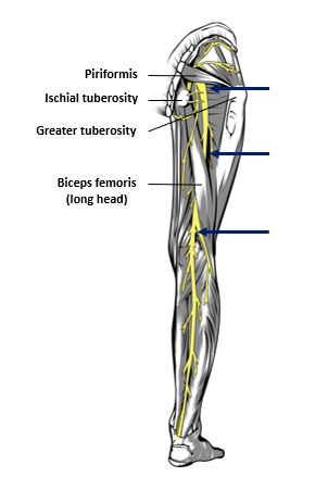
Branches of the sciatic nerve can become tethered to the scar following a hamstring injury, creating extraneural tension with or without local irritation(16). Peripheral sensitization can occur following sustained input to the hamstrings, resulting in local damage. Mounting clinical and MRI evidence support post-inflammatory scarring as a primary cause of irritation in this region (see figure 2).
The blue arrows indicate the common sites of irritation.
The sciatic nerve is stretched by an excursion of 28mm during a modified Straight Leg Raise with knee extension. However, the flexed, abducted, and externally rotated hip position can limit its excursion. Moreover, when clinicians flex the athlete’s hip to 90° while the knee is fully extended, the sciatic strain increases by 26%. Thus, impaired gliding of the sciatic nerve can disrupt its normal excursion and induce DGS(17). In addition, hamstring injuries reduce hamstring flexibility in the injured and uninjured limbs. This demonstrates the development of adverse neural tension and indicates abnormal physiological and mechanical responses produced by the nervous system(18).
Finally, any anatomic changes affecting how the femoral head sits in the acetabulum or other structural hip or pelvic abnormalities may result in subtle gait disorders that affect the hip, resulting in biomechanical pelvic abnormalities. Over time these abnormalities can contribute to the pathologic changes and clinical signs and symptoms of DGS and hamstring strains(18).
Conclusion
Hamstring injuries are the most common soft tissue injuries in sports. However, the cause may not always be the same. Therefore, clinicians must conduct a comprehensive neurodynamic assessment to ensure sciatic nerve integrity. Furthermore, following any injury around the posterior pelvic and thigh region, clinicians should include neurodynamic exercises to prevent secondary complications which may prolong rehabilitation. The posterior thigh remains a key performance area in most sports, and proper assessment and diagnosis are essential to keeping the athlete moving forward.
References
- Sports Med, 34(10), 681-695.
- Sports Med, 29(4), 273-287.
- Sports Health, 4(2), 107-114.
- Best Practice & Research Clin Rheumatology, 21(2), 261-277.
- European Spine J, 19(12), 2095-2109.
- Clinics in Sports Medicine 35(3) 469-486.
- Skeletal Radiology, 44(7), 919-934.
- The J of Bone and Joint Surgery. British volume, 93(10), 1300-1302.
- Acta Orthopædica Belgica, 76(3), 321-324.
- The American J of Sports Med 16, no. 5 (1988): 517-521.
- Best Practice & Research Clinical Rheumatology, 21(2), 261-277.
- Int J Sports Med. 2017;38(11):803–808.
- The Bone & Joint J, 102(5), 556-567.
- Int Orthop. 2017;41(7):1321–1328.
- J Bone Joint Surg Br. 2003;85-B(3):363–365.
- British J of General Practice, 69(687), 485-486.
- J of Sport and Health Science, 1(2), 92-101.
- Sports Med, 42(3), 209-226.
Newsletter Sign Up
Subscriber Testimonials
Dr. Alexandra Fandetti-Robin, Back & Body Chiropractic
Elspeth Cowell MSCh DpodM SRCh HCPC reg
William Hunter, Nuffield Health
Newsletter Sign Up
Coaches Testimonials
Dr. Alexandra Fandetti-Robin, Back & Body Chiropractic
Elspeth Cowell MSCh DpodM SRCh HCPC reg
William Hunter, Nuffield Health
Be at the leading edge of sports injury management
Our international team of qualified experts (see above) spend hours poring over scores of technical journals and medical papers that even the most interested professionals don't have time to read.
For 17 years, we've helped hard-working physiotherapists and sports professionals like you, overwhelmed by the vast amount of new research, bring science to their treatment. Sports Injury Bulletin is the ideal resource for practitioners too busy to cull through all the monthly journals to find meaningful and applicable studies.
*includes 3 coaching manuals
Get Inspired
All the latest techniques and approaches
Sports Injury Bulletin brings together a worldwide panel of experts – including physiotherapists, doctors, researchers and sports scientists. Together we deliver everything you need to help your clients avoid – or recover as quickly as possible from – injuries.
We strip away the scientific jargon and deliver you easy-to-follow training exercises, nutrition tips, psychological strategies and recovery programmes and exercises in plain English.


