You are viewing 1 of your 1 free articles
Fat facts for clinicians treating anterior knee pain

Injuries to the infra-patella fat pad (IFP) can be a cause of anterior knee pain in athletes – particularly in sports that require forced extension of the knee, as in kicking a ball or jumping. Understanding the anatomy of the IFP is important for the clinician, as the signs and symptoms of IFP injury are often misdiagnosed as patellofemoral pain or patellar tendinopathy.
IFP anatomy and function
The infra-patella fat pad or ‘Hoffa’s fat pad’, first described by Hoffa in 1904(1), is described as being intracapsular but extra synovial. It sits behind and around the patellar tendon and occupies space in the anterior knee joint(2,3). Some of the key anatomical features of the IFP are as follows (see figure 1):- Appears as a yellow structure and includes globules of fat that extend posteriorly and superiorly into the knee joint(4).
- The posterior surface blends with the synovial septum of the knee which is referred to as the ligamentum mucosum or the infrapatellar plica(3,5,6).
- The ligamentum mucosum (infrapatellar plica) extends posteriorly and connects to the intercondylar notch of the femur(4,6,7). In some knees, this ligamentum mucosum is continuous with the anterior cruciate ligament(4).
- Other known attachments of the IFP include(6,7):
- Proximal patella tendon
- Inferior pole of the patella
- Transverse meniscal ligament
- Medial and lateral meniscal horns
- Tibial periosteum
- At the edges of the IFP, the patellofemoral ligaments (Kaplan’s ligaments) appear as thickenings as they merge with the capsular synovium(8).
- The IFP has a rich blood supply via the genicular arteries and the dense innervation of nociceptive nerves make it susceptible to pain with impingement(9,10).
- Injections into the IFP may result in pain felt at the inferior patellar pole, deep to the patella, and sometimes medial thigh pain. They can also result in delayed activation of the VMO(11).
Figure 1: Structure of the IFP

Function
The IFP is a mobile and flexible structure, which can change position, deform, and produce volume changes during knee flexion and extension in order to adjust pressures at the knee(12,13). The primary function of the IFP is to reduce friction and shear between the tibia, femur, the patella, and the patellar tendon. Secondarily, it prevents pinching of the synovial membrane between the tibia and femur(14). It also improves the distribution of the lubricant effect of intra-articular joint fluid by effectively increasing the synovial surface(15,16).During knee flexion, the IFP pushes posteriorly and superiorly into the trochlear groove of the femur as the space between the patellar tendon and tibia decreases. The increase in pressure in the IFP during flexion helps stabilize the patella(16,17). During knee extension, the IFP moves anteriorly and creates space between the IFP and tibia and femur(12,13).
Diagnosis of IFP injury
The typical clinical presentation of patients with an IFP irritation include(8,18-20);- Anterior knee pain described as ‘burning and aching’.
- Location of pain that is inferior to the patella and on either side of the tendon.
- Tender and enlarged mass under the patella on palpation.
- Pain with forced full extension - either passively or active extension.
- Pain with prolonged flexion.
- A positive Hoffa’s test(21). Perform with the patient lying supine with the knee and hip flexed to 90 degrees. Press alongside the patellar tendon and ask the patient to extend the knee. If this causes pain it is deemed a positive ‘Hoffa sign’.
Imaging
The most commonly used imaging modality in IFP pathology is magnetic resonance imaging (MRI). Sagittal MRI is the best view to identify pathology. Fibrosis appears as a hypo-intense signal on T1-, T2- and proton density-weighted images(7,8,22), while edema within the IFP appears as a hyper-intense signal on T2-weighted images with fat saturation(7,15,23). Recent evidence also suggests dynamic ultrasound investigations are quite effective in identifying IFP pathology(24).Injury to the IFP
Injuries to the IFP can be broken down into subgroups.- Inflammation and fibrosis
Acute injury to the IFP due to direct trauma, repetitive microtrauma (eg excessive torsion), and iatrogenic injury lead to hypertrophy and fibrosis in the IFP. Blunt impact causing ACL rupture and patellar dislocation usually affects the IFP as well (7,19,23). Iatrogenic injury can occur after arthroscopy due to the portal placements(25). Symptoms become most apparent six months post-arthroscopy and slowly decreases over the next 6 months(26).
- Impingement of the IFP
Impingement at the anterior tibiofemoral joint or the lateral patellofemoral joint starts a cascade of inflammation and fibrosis(19,23,25). Adherence of the IFP to the anterior tibia is called ‘anterior interval scarring’(8), while impingement of the IFP may present with similar symptoms as other patellofemoral pathologies such as chondromalacia(14). Impingement is more common in women and the classic symptoms include anterior knee pain whilst going up and down stairs. Pre-existing ligamentous laxity that leads to knee hyperextension may also cause repetitive trauma and inflammation(21).
- Cyclops lesion
A nodular mass of soft tissue located in the posterior part of the IFP can form post ACL reconstruction anterior to the ligament graft. This leads to anterior knee pain and an inability to fully extend the knee(27).
- Synovitis
When the posterior layer of the IFP adheres to the synovial lining of the knee joint, repetitive stretching and irritation can lead to synovitis and cause anterior knee pain(17).
- Neoplasms
Although rare, benign tumors and synovial sarcomas have been described, and present as anterior knee pain(28).
Conservative management
The goal of conservative intervention is to restore normal patellofemoral joint biomechanics via VMO retraining; stretching tight tissues implicated in patellofemoral malalignment (eg lateral quadriceps and distal ITB); correcting hip biomechanics that promote hip internal rotation and valgus knee collapse; and taping the IFP(29).*Taping
The purpose of taping the IFP is to draw the inferior pole of the patella away from the symptomatic IFP. Shown below is a taping procedure that is suitable for athletes, where flexion loads are being imposed on the knee (the classic procedure often results in the tape being pulled away from the skin during exercise). This ‘athletic’ taping procedure is described and shown in the sequence of photos below:
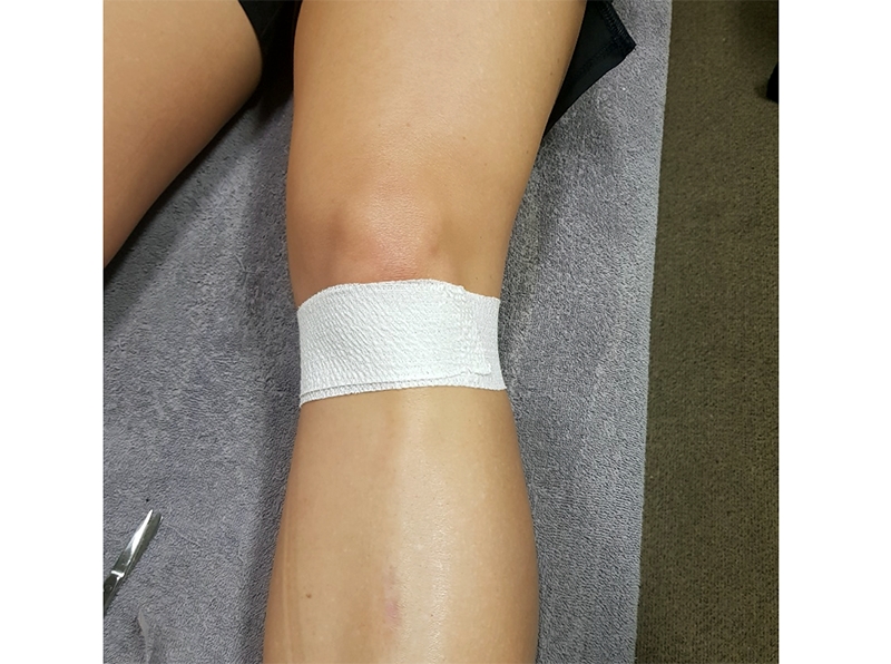
- Use a tearable or non-tearable EAB type tape. Start around the posterior aspect of the fibula head and wrap anteriorly around the patellar tendon. Continues posteriorly to completely encircle the knee (this is placed on lightly so as to not occlude blood flow) (see figure 2).
- Follow with a rigid 38mm tape starting behind the fibular head and held on length between one finger (see figure 3)- then placed posteriorly on the medial posterior knee. Create a fold in the tape (see figure 4).
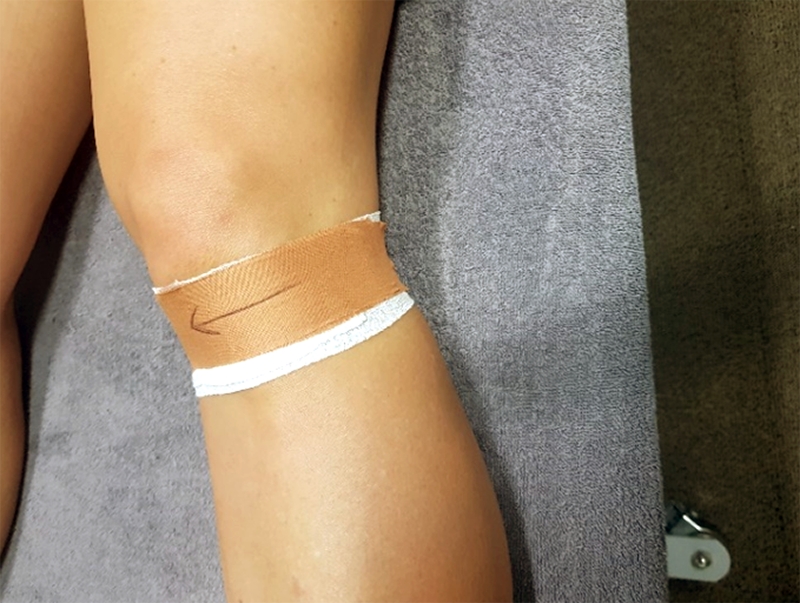
- Use a second short strip of 38mm rigid tape to pull the fold of tape medially. This draws the patellar tendon, the IFP and the patella medially and creates space away from the IFP (see figure 5).
Figure 2: Step 1 base layer of tape
Figures 3 and 4: Step 2 creating an anchor and fold in tape
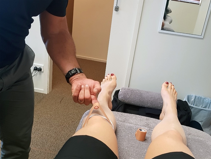
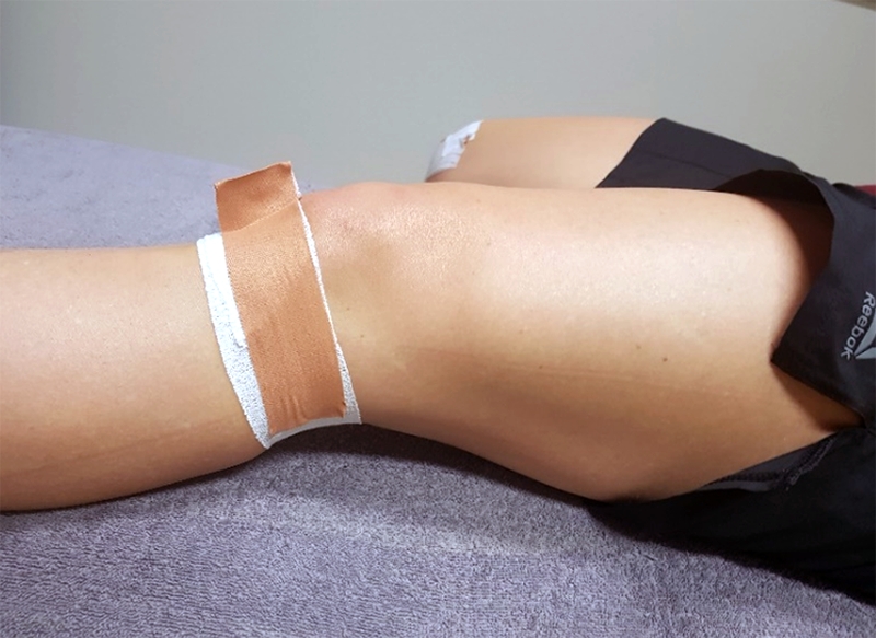
Figure 5: Step 3 Drawing structures medially
*VMO retraining
Have the athlet perform quadriceps exercises that focus on VMO activation away from positions of full lock out extension as those may irritate the IFP. Closed kinetic-chain exercises are usually preferred as they allow control of the knee to avoid full extension. Open kinetic-chain exercises are usually a poor choice as they may encourage forced hyperextension(30). The ‘door jam’ is an example of closed kinetic chain exercise that only uses the 0-30 degrees angle of flexion (see figure 6).
Figure 6: "Door jam" exercise
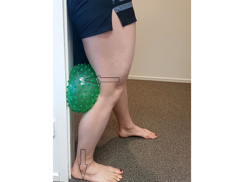
The ‘door jam’
- Use an inflatable ball that is able to deform and compress. A rigid ball such as medicine ball or soccer ball will not work.
- Place the ball just above the knee crease so the bulk of the ball is against the hamstring muscle and calf muscles.
- Forcefully push the heel of the foot into the ground.
- Actively push the thigh backwards into the ball (to create knee extension).
- The ball will compress; however it will still prevent the knee from fully extending and thus protecting the IFP.
- Hold this for 5-10 seconds and repeat for the desired number of repetitions.
*Gluteal retraining
The purpose of activation work on the posterior fibres of gluteus medius and deep external rotators is to prevent the hip from internally rotating during stance phase – ie to reduce the malalignment issues around the patellofemoral joint. These are preferably done in weight bearing positions and involve the triple extension phase of walking and running.
Conclusion
Injuries to the IFP may mimic other anterior knee pathologies such as chondromalacia patella, patella tendon pathology, patellofemoral maltracking and anterior meniscal pathologies. IFP injury is more common in sports that require forced extension (kicking), hyper-extension (gymnastics), and loaded jumping sports (basketball and volleyball). It is reasonably well managed conservatively in the athletic population through rehabilitative corrective exercise and taping.References
- JAMA 1904; 43: 795-6
- Clin Sports Med 2002; 21 (3): 335-47
- Am J SportsMed 2004; 32 (8): 1873-80
- Arthroscopy 2009; 25 (8): 839-45
- Knee Surg Sports Traumatol Arthrosc 2014; 22(2):247–262
- Knee Surg Sports Traumatol Arthrosc 2005; 13 (4): 268-72
- Radiographics 1997; 17 (3): 675-91
- Am J Sports Med 2008; 36 (9): 1763-9
- Arch Orthop Trauma Surg 1995; 114 (2): 72-5
- Am J Sports Med 1998; 26 (6): 773-7
- Arthritis Rheum 2009; 61 (1): 70-7
- Clin Biomech 2010; 25 (5): 433-7
- Clin Anat 1992; 5 (2): 107-12
- Insights Imaging 2016: 7:373–383
- Skeletal Radiol 2004; 33 (8): 433-44
- Boshnack Knee Surgery Sports Trauma Arthroscopy 2005 13(2). 135-141
- J Arthroplasty 2003; 18 (7): 897-902
- Knee 1997; 4 (4): 227-36
- Arthroscopy 2007; 23 (11): 1180,1186.e1
- J Knee Surg 1988; 1:134-9
- Diagn Interv Imaging 2014;95:1079—84.
- Am J Sports Med 1992; 20 (5): 519-25; discussion 525-6
- Arch Orthop Trauma Surg 2010; 130 (8): 1041-51
- The Knee 2018;25: 279–285
- Arthroscopy 1994; 10 (2): 184-7
- Radiat Med 2000; 18 (1): 1-5
- AJR Am J Roentgenol 174(3):719–726
- International Orthopaedics (SICOT) (2013) 37:2225–2229
- Sports Med 2012; 42 (1): 51-67
- Sport Health 1991; 9 (4): 7-9
Newsletter Sign Up
Subscriber Testimonials
Dr. Alexandra Fandetti-Robin, Back & Body Chiropractic
Elspeth Cowell MSCh DpodM SRCh HCPC reg
William Hunter, Nuffield Health
Newsletter Sign Up
Coaches Testimonials
Dr. Alexandra Fandetti-Robin, Back & Body Chiropractic
Elspeth Cowell MSCh DpodM SRCh HCPC reg
William Hunter, Nuffield Health
Be at the leading edge of sports injury management
Our international team of qualified experts (see above) spend hours poring over scores of technical journals and medical papers that even the most interested professionals don't have time to read.
For 17 years, we've helped hard-working physiotherapists and sports professionals like you, overwhelmed by the vast amount of new research, bring science to their treatment. Sports Injury Bulletin is the ideal resource for practitioners too busy to cull through all the monthly journals to find meaningful and applicable studies.
*includes 3 coaching manuals
Get Inspired
All the latest techniques and approaches
Sports Injury Bulletin brings together a worldwide panel of experts – including physiotherapists, doctors, researchers and sports scientists. Together we deliver everything you need to help your clients avoid – or recover as quickly as possible from – injuries.
We strip away the scientific jargon and deliver you easy-to-follow training exercises, nutrition tips, psychological strategies and recovery programmes and exercises in plain English.


