Assessing and managing acetabular labral tears
Acetabular labral tears of the hip are one of the more challenging injuries for clinicians to diagnose and manage. Trevor Langford looks at what the recent evidence says.

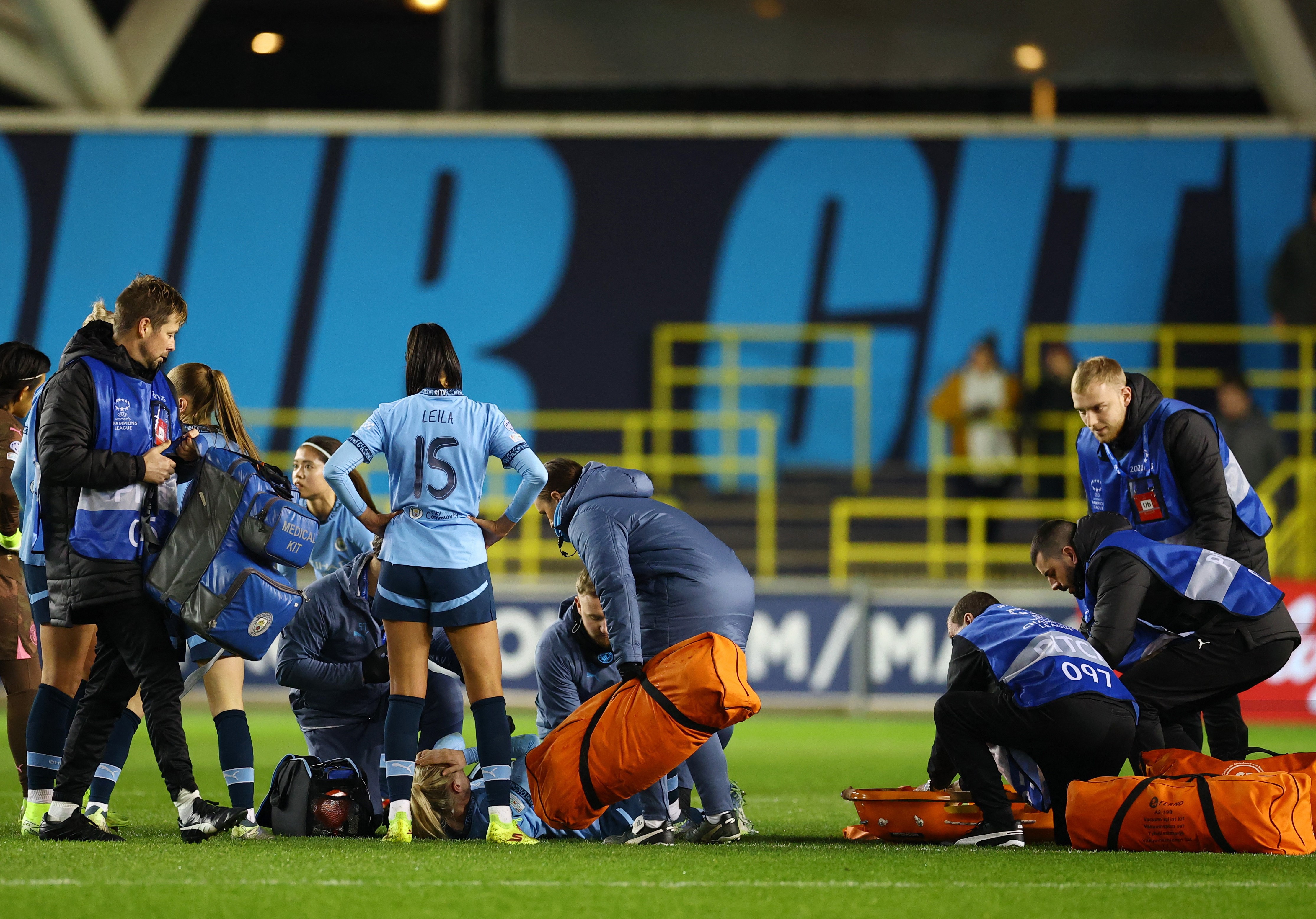
England’s Ruben Loftus-Cheek holds his hip, 2017
Mechanical disruption of the hip joint is often related to an acetabular labral tear (ALT) and can be associated with intraarticular snapping hip syndrome in up to 80% of cases(1). Labral tears affecting the hip joint are prevalent in 22-55% of patients with hip or groin pain and evidence suggests that an untreated ALT may predispose an individual to early degenerative hip arthritis, which has created a widespread interest within clinical practice and the literature(2,3).
The examination and imaging techniques with suspected ALT’s have increased greatly in the past decade, but an assessment of a labral tear still remains complex. The purpose of this article therefore is to review the anatomy and biomechanics of the acetabular labrum, the examination techniques and the treatment and management options available.
Anatomy and biomechanics
The labrum increases the surface area of the acetabulum by 22% and the volume by 33% and it functions by forming a seal for the head of femur to rotate in (see figure 1). From a cross-sectional view, the labrum is triangular in its appearance with the extra articular zone being dense connective tissue that has a rich blood supply and the intra articular zone largely having no blood supply(3).
At the extreme end ranges of hip motion, the labrum is stressed by compressive forces -a tear at this point can affect joint stability and load distribution(2). Furthermore, pain receptors have been located in the superior and anterior areas, and the labrum is therefore considered to be a pain-generating structure(4). It is at the anterior surface where an ALT is most at risk due to the compressive forces at the end point of hip flexion (see figure 1)(5). Also of note is that structural abnormalities such as retroverted acetabulum and coxa valga have been observed concurrently in patients (87%) with labral tears(6).

Figure 1: illustration of an acetabular labral tear at the anterior surface
Examination and assessment
An ALT is complex to diagnose and despite recent advances in medical imaging and assessment techniques, one report identified that on average, patients visited three healthcare providers and also waited for 21 months before an ALT was correctly diagnosed(3). When examining a patient with a suspected ALT, clinicians should also consider femoro-acetabular impingement (FAI) and acetabular cartilage damage, and MRI imaging should be used to support the clinical findings(3).
Acetabular labral tears are often the result of cutting, pivoting, twisting as well as repetitive movements into end range hip flexion commonly found in tennis players, footballers and runners. This might help explain why researchers from the New England Baptist hospital in Boston, USA found that from 436 arthroscopies of labral tears in athletes, 273 (62%) also had articular cartilage damage(6). However the exact mechanism of an ALT injury may not always be apparent to the patient, as it may be degenerative, congenital or traumatic in its occurrence(2).
During a physical assessment of a hip related injury it is essential to be vigilant for a non-musculoskeletal related pathology. Hip related pain may be associated to an ALT but may also be the result of lumbar spine or pelvic girdle dysfunction, abdominal viscera and the peripheral nervous system(2). Pain at rest, night pain, fever, night sweats, and generally feeling unwell and unexplained weight loss are indicators of a non musculoskeletal pathology and require referral for further examination by a healthcare provider(2). Reiman and colleagues also indicated that hip pain may be related to the abdominal and pelvic organs and a musculoskeletal injury must not be assumed(2).
A patient with an undiagnosed ALT may also present with synovitis and joint inflammation and may adopt positions of hip flexion, external rotation and slight abduction, which place the capsule at its largest potential volume to reduce the stress on the labrum. Positions which include flexion and or adduction have been found to increase the overall load on the labrum and this are consciously avoided.
The combined impingement position of flexion, adduction and internal rotation, known as ‘FADDIRs test’ increases stress to the labrum, but is also a contributor to intra-articular hip pathology(7). Patients with an ALT may also complain of pain on squatting, stepping up with the involved limb, or sitting in a chair with the hips positioned lower than the knees. In addition a patient with an ALT is unlikely to extend fully at the hip during gait as this places the greatest load to the anterior joint capsule and consequently stress to the anterior labrum (see table 1).
Table 1: Specific tests for acetabular labral tear
| Physical test | Testing procedure | Positive result indicating a possible labral tear |
| Thomas Test | The patient sits at the edge of the plinth and lies on their back with both knees to the chest. One knee (the uninvolved side) is held to the chest and the involved limb is slowly lowered into extension of the hip by the clinician. The knee is allowed to extend. The patient is instructed to pull his or her pelvis into posterior rotation. The clinician may use a goniometer to measure the extension angle of the hip and/or the knee. | The patient’s groin or hip pain is reproduced with or without a click. |
| FADDIR Test | The patient is positioned in supine. The therapist passively moves the patient’s leg to approximately 90 degrees of hip and knee flexion. The leg is then passively adducted and internally rotated at the hip. | The patient’s groin pain is reproduced. |
| Flexion-internalrotation test | The patient is supine. The clinician passively performs the combined movements of flexion to 90 degrees and internal rotation of the hip is carried out. | The patient’s groin pain is reproduced. |
| Scour test | The patient starts in a supine position. The clinician flexes the patient’s hip and knee, performing a sweeping motion from external to internal rotation as an axial load is applied. | Reproduction of the patient’s pain or apprehension at a specific point. |
| Internal rotationwith overpressuretest | The patient is supine. The clinician passively moves the hip to 90 degrees flexion followed by internal rotation with overpressure at the end of range. | Pain or discomfort reproduced in the groin. |
| Resisted straightleg raise test | The patient lies supine with both legs extended, and with the trunk supported upright with both arms. The patient raises their leg 30 cm off the table with the clinician applying a downward force at the distal thigh with the patient aiming to resist this force. | Reproduction of pain in the lower quadrant or anterior hip. |
| Internal rotationflexion-axialcompression test | The patient lies supine. The clinician passively performs the combined motions of hip internal rotation, flexion and axial compression down into the acetabulum (longitudinally through the femur). | Pain or discomfort reproduced in the groin. |
| Postero-inferiorlabrum test | The patient lies supine, close to the edge of the table. The clinician passively moves the hip into hyperextension, abduction and external rotation. | Reproduction of discomfort and apprehension |
Surgical versus non-surgical treatment
Hip arthroscopy is a widely used treatment adjunct in patients presenting with an ALT symptomatic of longer than four weeks and confirmed by MRI (magnetic resonance imaging) or MRA (magnetic resonance arthrogram)(4). Hip athroscopy for an ALT may include either labral debridement or labral repair. In contrast to surgical repair there is limited support for conservative treatments for an ALT. However researchers from Sao Paulo, Brazil have provided a case series of four patients that underwent a rehabilitation programme for an ALT without surgery(8).
The four patients were diagnosed by an MRI scan and underwent a 3-phase programme with the first being pain control, trunk and hip stabilisation, re-education and correction of abnormal joint movement. Phase two focused on restoring normal range of motion, muscular strength and commencing sensory motor training. The final phase of their rehabilitation programme focused on preparing the athlete for a return to sport. The four patients involved in the case series were in their mid twenties and were from both sedentary and athlet ic backgrounds(8). The results of the conservative rehabilitation programme yielded a decrease in pain levels, functional improvement and correction of muscle imbalances. Increased muscle strength was noted with the hip flexors increasing from 1% to 39%, hip abductors increasing from 18% to 56% and the hip extensors increasing from 68% to 139%. The strength of this research is limited, with the case series being just four patients but nevertheless could provide a very good proactive approach whilst a patient is awaiting an arthroscopy.
Table 2: Suggested exercises to include during weeks 1-4 post surgical intervention
| Ankle Range of movement exercises |
| Gluteal, quadriceps, hamstring, transverses abdominis isometric contractions |
| Passive range of movement exercises |
| Heel slides |
| Walking in water (once sutures have healed), progress to jogging with flotation device within hip limits |
| Cycling with no resistance |
| Side clam exercises progressing to side lying hip abduction (labral debridement only) |
Rehabilitation
Rehabilitation following surgical repair of an ALT is limited in terms of its evidence, both within the surgeons own rehabilitation protocol and the therapist’s expertise.
Phase 1 (weeks 1–4)
- Following labral debridement, weight bearing should be limited to 50% partial weight bearing for 7-10 days, with flexion limited to 90° for 14 days. A labral debridement also has no limits postoperatively into abduction, internal or external rotation or extension. In contrast a labral repair should maintain non-weight bearing or toe-touch weight bearing for three to six weeks post operatively.
- The ranges of movement should be far more conservative in labral repair, while internal and external rotation should be conservatively moved into for 3 weeks. Do note that if other procedures are carried out such as microfracture repair then the post-op protocol may well be different.
- During the immediate post-operative period, it is essential to manage pain, reduce swelling and initiate early movement at the affected limb, but it is also essential to focus on other factors such as core activation and abductor control (see table 2). A strong association exists between decreased activity of the hip abductor muscles and lower kinetic chain injuries, including anterior knee pain(9). Therefore, once the hip ranges are accessible, it is essential to encourage a patient to activate the deep hip and trunk stability muscles to prevent secondary injuries from occurring.
Phase 2 (weeks 5–7)
- During this stage of rehabilitation it is essential to restore normal range of movement with an emphasis on increasing strength and developing flexibility of the muscles crossing the hip joint.
Exercises to include during weeks 5-7

- Double leg standing squats with use of a swiss ball
- Cycling with increased resistance

- Seated resistance internal / external rotation

- Double or single leg bridging

- Kneeling hip flexor stretches

- Side stepping with clini band around knees
- Crosstrainer
Phase 3 (weeks 8-12)
- This phase is a great opportunity to develop cardiovascular fitness and functional control of the hip. Functional stability exercises should be carried out in a standing position with an emphasis on maintaining and improving stability ready for sporting participation. Exercises to include in this phase are walking lunges, lunges with trunk rotation over the front leg and a Swiss ball programme appropriate for challenging the core muscles. Garrison and colleagues have provided criteria (see table 3) to follow ready for progression to phase four which is preparation for return to sport(10).
Phase 4 (weeks 12+)
- This phase of the rehabilitation programme is the time to be preparing the athlete for their return to sport, and particularly their position it has specific tasks. If the athlete is a defender in football, they should be replicating tasks specific to their position. Prior to the patient resuming full training, they must be able to demonstrate the same neuromuscular control as the uninvolved side.
Table 3: Training for fitness and functional control of hip
| Symmetrical hip ranges of motion between left and right legs |
| Symmetrical flexibility of the psoas and piriformis muscles |
| No Trendelenburg sign of either side indicating appropriate hip abductor control |
Summary
A suspected ALT using the history and clinical texts should be confirmed using an MRI or MRA to confirm the presence of an ALT but also to exclude any referred pain masquerading as a musculoskeletal injury. An appropriate rehabilitation programme should be started immediately to improve hip and trunk control and to manage pain. This will allow the patient to proceed through the surgery with greater ease having already commenced a rehabilitation programme.
You need to be logged in to continue reading.
Please register for limited access or take a 30-day risk-free trial of Sports Injury Bulletin to experience the full benefits of a subscription. TAKE A RISK-FREE TRIAL
Newsletter Sign Up
Subscriber Testimonials
Dr. Alexandra Fandetti-Robin, Back & Body Chiropractic
Elspeth Cowell MSCh DpodM SRCh HCPC reg
William Hunter, Nuffield Health
Newsletter Sign Up
Coaches Testimonials
Dr. Alexandra Fandetti-Robin, Back & Body Chiropractic
Elspeth Cowell MSCh DpodM SRCh HCPC reg
William Hunter, Nuffield Health
Be at the leading edge of sports injury management
Our international team of qualified experts (see above) spend hours poring over scores of technical journals and medical papers that even the most interested professionals don't have time to read.
For 17 years, we've helped hard-working physiotherapists and sports professionals like you, overwhelmed by the vast amount of new research, bring science to their treatment. Sports Injury Bulletin is the ideal resource for practitioners too busy to cull through all the monthly journals to find meaningful and applicable studies.
*includes 3 coaching manuals
Get Inspired
All the latest techniques and approaches
Sports Injury Bulletin brings together a worldwide panel of experts – including physiotherapists, doctors, researchers and sports scientists. Together we deliver everything you need to help your clients avoid – or recover as quickly as possible from – injuries.
We strip away the scientific jargon and deliver you easy-to-follow training exercises, nutrition tips, psychological strategies and recovery programmes and exercises in plain English.


