Achilles Tendon Rehabilitation: Maximize Anabolic Tendon Remodeling
Unlock the secrets of Achilles tendon rehabilitation with Renée Da Silva, as she reveals crucial insights for clinicians to optimize exercise selection and achieve anabolic remodeling.
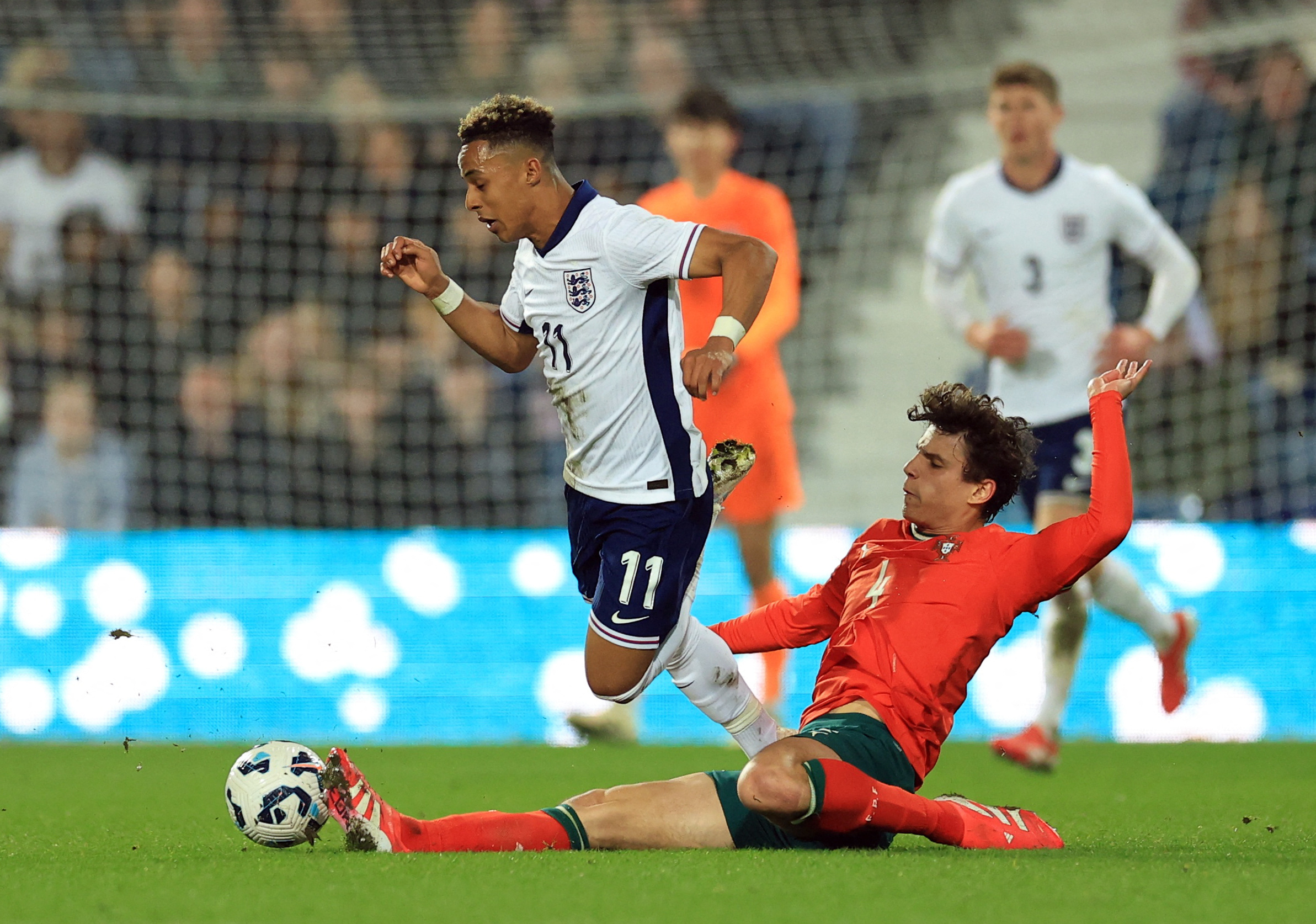
LA Clippers guard Russell Westbrook drives around Oklahoma City Thunder guard Shai Gilgeous-Alexander during the second half at Paycom Center. Mandatory Credit: Alonzo Adams-USA TODAY Sports.
Tendons, like other musculoskeletal tissues, are responsive to their mechanical environment, with too much mechanical stimulation leading to tissue damage and too little causing tissue atrophy(1,2). The human Achilles tendon is a complex three-dimensional structure that enhances power production and efficiency of the triceps surae muscle-tendon complex during movement(3-6).
Biomechanics, mechanobiology, and adaptation
Musculoskeletal tissues are sensitive to their mechanical environment, with excessive and insufficient loading resulting in reduced tissue strength(7,8). Whereas bone appears to be most sensitive to loading at high strain rates, tendons are particularly sensitive to the tissue strain magnitude(9-14).
Strain magnitude is a key control variable for tendon adaptation. Therefore, clinicians must ensure adequate loading when designing exercise and rehabilitation programs. The efficacy improves when the strain magnitude experienced by the Achilles tendon during exercise is in the anabolic range of approximately 5–6%. In this range, tendon remodeling exceeds strain-induced tendon damage(15).
To measure the tendon strain effectively, researchers attach an ultrasound probe to the leg to track the motion of the gastrocnemius muscle tendon junction to estimate the change in tendon length relative to a motion capture marker placed on the calcaneus(16,17).
Achilles Tendon Strain
Exercise-based interventions that target strains (i.e., strain magnitude, strain rate, and volume) associated with maximum anabolic tendon adaptation improve outcomes compared with non-targeted exercise-based interventions(1). Furthermore, strain and not force (or stress) is the primary mechanical stimulus that drives tendon remodeling(18-21). Researchers and clinicians must establish approaches to quantify tendon strain during training and rehabilitation(1,2).
Researchers in Australia set out to estimate in vivo free Achilles tendon strain during selected rehabilitation, locomotor, jumping, and landing tasks. They wanted to better understand the strains the Achilles tendon experienced during commonly prescribed exercises and locomotor tasks. The study results demonstrate that the average and peak free Achilles tendon strains are significantly higher in hop landing and running tasks than heel rise. Moreover, the average and peak free Achilles tendon strains are significantly lower in walking than heel rise. The researchers found that the average strain was significantly lower during the countermovement jump push-off phase, and the peak strain was significantly higher than the heel rise (see figure 1).
Once the researchers added weight (20% body weight) to the heel-rise, the average and peak free Achilles tendon strains were significantly higher than unloaded repetitions. Notably, running speed increases the average and peak free Achilles tendon strains (five m/s compared with three m/s)(22).
The study results also demonstrate that clinicians must introduce plyometric exercises into the rehabilitation program once athletes have the necessary capacity. The results clearly show that plyometric tasks exceed the peak force of heel raises (with or without added weight). Therefore, it would be unreasonable to allow an athlete to return to sport without adequate exposure to sport-specific, plyometric tasks.
As tendon strain is the primary mechanical stimulus that drives tendon remodeling, the study results provide clinicians with guidelines for exercise selection to maximize anabolic tendon remodeling during training and rehabilitation.
You need to be logged in to continue reading.
Please register for limited access or take a 30-day risk-free trial of Sports Injury Bulletin to experience the full benefits of a subscription. TAKE A RISK-FREE TRIAL
Newsletter Sign Up
Subscriber Testimonials
Dr. Alexandra Fandetti-Robin, Back & Body Chiropractic
Elspeth Cowell MSCh DpodM SRCh HCPC reg
William Hunter, Nuffield Health
Newsletter Sign Up
Coaches Testimonials
Dr. Alexandra Fandetti-Robin, Back & Body Chiropractic
Elspeth Cowell MSCh DpodM SRCh HCPC reg
William Hunter, Nuffield Health
Be at the leading edge of sports injury management
Our international team of qualified experts (see above) spend hours poring over scores of technical journals and medical papers that even the most interested professionals don't have time to read.
For 17 years, we've helped hard-working physiotherapists and sports professionals like you, overwhelmed by the vast amount of new research, bring science to their treatment. Sports Injury Bulletin is the ideal resource for practitioners too busy to cull through all the monthly journals to find meaningful and applicable studies.
*includes 3 coaching manuals
Get Inspired
All the latest techniques and approaches
Sports Injury Bulletin brings together a worldwide panel of experts – including physiotherapists, doctors, researchers and sports scientists. Together we deliver everything you need to help your clients avoid – or recover as quickly as possible from – injuries.
We strip away the scientific jargon and deliver you easy-to-follow training exercises, nutrition tips, psychological strategies and recovery programmes and exercises in plain English.


