Sinus tarsi syndrome: a possible source of lateral ankle pain

Sinus tarsi syndrome (STS) is a frequently misdiagnosed condition in which patients have pain over the lateral aspect of the ankle (the sinus tarsi region) with an ‘unstable’ sensation in the rearfoot. STS sufferers usually have a history of an inversion ankle injury, which may damage the usual lateral ankle ligaments, leading to chronic pain in and around the sinus tarsi weeks to months after the injury.
Anatomy and biomechanics of sinus tarsi
The sinus tarsi has been compared with the intercondylar fossa of the knee(1), and forms part of the subtalar joint complex. The sinus tarsi is the opening of the talocalcaneal sulcus, which is shaped like a long funnel. It has a large anterolateral opening, narrowing down to a smaller posteromedial opening. The anterior opening is found anterior and inferior to the lateral malleolus, while the posterior opening sits behind the sustentaculum tali below the medial malleolus. The inside of the sinus tarsi tunnel is filled with a host of numerous structures (see figure 1). These include:- The large interosseous talocalcaneal ligament (ITCL)- The medial band of the ITCL is a single fibrous band. The lateral half splits into two bands, which are separated by a vascular and innervated adipose tissue. The ITCL is taut in supination and relaxed in pronation.
- Adipose tissue- Serves as a bedding for numerous mechanoreceptors and free nerve endings, which along with the ligaments and muscles provide proprioceptive information on the movement of the foot and ankle.
- The inferior part of the extensor retinaculum (IER) -This lies over the lateral opening and serves as a covering to the sinus tarsi.
- The extensor digitorum brevis muscle- Attaches to the medial and distal aspect of the sinus tarsi, running over the calcaneocuboid joint towards the toes.
- The cervical ligament (CL).
- A synovial membrane– Surrounds the sinus tarsi
Figure 1: Anatomy of the sinus tarsi

The main ligament is the ITCL. This is a wide and very strong ligament that originates from a broad attachment in the middle of the canal on the surface of the calcaneus and runs anteromedially to the deepest portion of the tarsal canal, where it inserts on the talus. The CL is a smaller band, which has its origin on the lateral calcaneus, just medial to the attachment of the extensor retinaculum of the foot, and passes medially through the centre of the canal as it inserts on the talus(2).
A recent study published described new findings on the anatomical relationship between the capsules and each ligamentous structure of the subtalar joint(3). It demonstrated (in a cadaveric study) that the tarsal canal and sinus consisted of three structured layers:
1) The anterior capsule of the posterior talocalcaneal joint, including the anterior capsular ligament.
2) The interosseous talocalcaneal ligament (ITCL) and inferior extensor retinaculum (IER) layers
3) The posterior capsule of the talocalcaneonavicular joint, including the cervical ligament (CL).
The tarsal canal ligaments maintain alignment between the talus and calcaneus and limit inversion. The main stabilising ligament of the lateral ankle is the calcaneofibular ligament. In addition, the ITCL is taut when the foot is supinated, and the CL helps resist hindfoot varus forces. With inversion trauma, the ligaments are usually injured in the following order: anterior talofibular ligament (ATFL), calcaneofibular ligament (CFL), CL, and ITCL. The more severe the injury, the more of these ligaments are injured. Thus, tarsal canal ligament injury never occurs as an isolated lesion with an inversion sprain; it will also be associated with damage to the ATFL and/or the CFL(1,4).
Pathogenesis of STS
The syndrome known as STS was first described by O’Connor in 1958(5)and the true incidence of STS is unknown. STS has been associated with ankle sprains that may also result in talocrural joint instability(1). Some key findings between the correlation of ankle sprain and STS include:- 10-25% of patients with chronic talocrural joint instability will also have subtalar joint instability(6).
- Nine out of twelve patients with recurrent ankle sprains had signs of increased talocrural and subtalar joint motions upon a radiographic examination(7).
- Previous inversion sprain has been found in 70% of STS sufferers(8-10).
Due to the abundance of synovial tissue, the sinus tarsi is prone to synovitis and inflammation when injured. Injury to the sinus tarsi falls into three broad categories:
- An inversion injury to the ankle that also injures the sinus tarsi. These injuries cause instability of the subtalar joint resulting in excessive supination and pronation movements. The excessive movement of the subtalar joint imparts increased forces onto the synovium of the subtalar joint and across the sinus tarsi tissues. The excessive forces result in subtalar joint synovitis with chronic inflammation and infiltration of fibrotic tissues in the sinus tarsi, which are responsible for the characteristic anterolateral ankle pain of STS(11).
- Chronic inflammation due to excessive bouts of pronation.
- Repeated forced eversion under impact load such as jumping. This mechanism is thought to create a ‘whiplash injury’ to the rearfoot, with the talus moving anteriorly over the calcaneus(6).
Signs and symptoms
Because the pain usually presents months after an injury to the lateral ankle ligaments, STS is often misdiagnosed, and STS may often simply be confused with chronic ankle instability. Some of the features that may alert the practitioner that the patient has a STS include(1-5):- Vague and poorly localised anterolateral ankle pain. Because the synovitis and fibrotic tissues associated with STS will take time to develop, athletes with injuries to the subtalar joint may not initially have symptoms that can be localised to the sinus tarsi.
- Morning pain that usually subsides with use.
- Pain running on a curve in the direction of the affected ankle.
- Feeling of ankle and foot stiffness.
- Feeling of instability and weakness of the ankle. Athletes involved with cutting and jumping activities on firm surfaces will have the greatest difficulty with subtalar instability as these activities will cause excessive movements of the subtalar joint to the end ranges of pronation and supination.
- Difficulty walking on uneven ground.
- Pain on palpation and localised swelling over the sinus tarsi opening in the anterolateral ankle.
- Normal talocrural movement but stiff subtalar joint movements.
- Pain of forced eversion of the subtalar joint.
Physical examination
There is no definitive test for STS. However, stability of the subtalar joint may be assessed with medial and lateral subtalar joint glides performed by moving the calcaneus over a stabilised talus in the transverse plane, and with subtalar joint distraction (7,10). One test has been described by Therman et al (1998), which aims to assess stability of the subtalar joint(12). This is performed as follows:- The test is performed with the athlete in supine, and with the ankle in 10 degrees of dorsiflexion to keep the talocrural joint in a stable position.
- The forefoot is first stabilised by the examiners hand, while an inversion and internal rotational force is applied to the calcaneus.
- An inversion force is applied to the forefoot.
- The examiner assesses for an excessive medial shift of the calcaneus and a reproduction of the athlete’s complaint of instability and symptoms.
Reproduction of the athletes feeling of instability or giving way may be reproduced by having the athlete single leg stand on the affected side and perform rotating motions of the leg and foot, which also produces their symptoms. The definitive diagnosis is usually an injection of local anaesthetic into the sinus tarsi to eliminate pain with movement and testing(13).
Imaging and diagnosis
Plain film X-ray does not provide a great deal of information regarding injury to the sinus tarsi. Radiographs of the subtalar joint are usually performed with Broden stress views, which are a series of oblique-lateral views performed with the ankle and foot placed in inverted and supinated positions.Magnetic resonance imaging (MRI) is the best method to visualise the structure within the sinus tarsi, especially the ITCL and CL(11). The most distinct finding for individuals with STS is a bright signal seen on T2-weighted images found in the area for sinus tarsal adipose tissue; this represents an infiltration or replacement of this tissue with inflammatory cells and fibrotic tissue(14). The MRI findings may also show degenerative changes in the subtalar joint(15). However, the gold standard for any intra-articular injury, including injury to the sinus tarsi, is arthroscopic investigation(16):
Treatment
Managing STS usually begins with conservative measures prior to surgical consideration. Treatment may include:- Relative rest.
- NSAID medication.
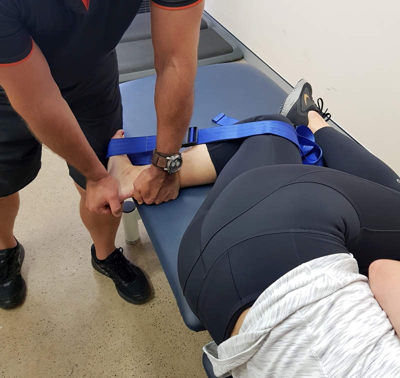
- Joint mobilisation of the subtalar joint and talocrural joint (see figure 2).
- Proprioception retraining.
- Soft tissue work and dry needling to muscles such as triceps surae, tibialis posterior/anterior and peroneals, which may help with compressive forces(17).
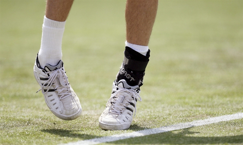
- Taping or strapping to limit movements of the subtalar joint and the midfoot. Viczenzio et al (2005)(18)have described a modified ‘low-dye’ taping method that uses a calcaneal sling, intended to provide support to the medial longitudinal arch of the foot (see figure 3). This method could be used to control or reduce the amount of pronation through the subtalar joint during walking and running activities.
- Footwear and orthotics for overpronation.
- Direct injection of a corticosteroid into the sinus tarsi(19, 20).
Figure 2: Mobilisation of the subtalar joint and talocrural joint

Subtalar joint glide: Position the patient in side lying with affected side down, Bend the knee to 100 degrees, placing the foot over the edge of the bed. Place a seatbelt looped around the lower thigh and around the midfoot and tighten the belt so that the ankle is locked into dorsiflexion. Place one hand under the fibula to protect the fibula from the edge of the bed while the other hand grasps the medial calcaneus and apply a gentle downward force to the calcaneus. This will glide the calcaneus laterally in relation to the talus.
Figure 3: Taping or strapping to limit movements of the subtalar joint and the midfoot

Surgery
Athletes who fail conservative management may need arthroscopic exploration and reconstruction of the subtalar joint in order to return to their athletic pursuits. The options available that have been described in the literature include:- Synovectomy of the subtalar joint, along with an arthrotomy of the subtalar joint, can be used to remove chronic synovitis and arthrofibrosis, which is commonly found in STS(21).
- Tarsectomy involves excision of the tarsal canal contents, including the fat plug, nerves, damaged capsule, and, occasionally, damaged ligaments. Sinus tarsectomy has an excellent cure rate(1,20).
- Surgical reconstructions of the ITCL and CL are made by splitting the tendon of the peroneus brevis, and routing the graft through bone tunnels made through the calcaneus and the talus(22).
- For recalcitrant cases and severe degeneration, triple arthrodesis of the ankle (fusion of the subtalar, calcaneocuboid, and talonavicular joints) may be the only surgical solution. However, this option is surgery of last resort(23).
Athletes who undergo ligamentous reconstructions are ususally immobilized for a 6-week period, followed by a rehabilitation program to regain normal ankle mobility, strength, and balance. Return to athletic activities usually begins at 4-6 months post-operatively(22). Common post-operative problems are transient loss of sensation of the lateral ankle and foot and persistent peroneal weakness(24).
References
- International Orthopaedics. 1981; 5 : 117-130
- Magn Reson Imaging Clin N Am. 1994;2(1):59-65
- Foot & Ankle International. 2018; 39(11). 1360 –136
- J Foot Surg. 1989;28(1):3-6
- J Bone Joint Surg (Am) 1958;40(3):720-726
- Foot Ankle Clinics. 2002;7: 577-609
- Med Sci Sports Exerc. 1999;31:1501-1508
- Arthroscopy 1998. 14; 373-381
- Foot and Ankle Int. 1998; 19. 462-465
- Sports Med. 2006;36:263-277
- Clin Pod Med Surg. 2005;22:63-77
- Foot Ankle. 1997; 18:163-169
- Renstrom PA, Kannus P: Sinus tarsi syndrome, in Delee JC, Drez D Jr (eds): Orthopaedic Sports Medicine: Principles and Practice. Philadelphia, WB Saunders, 1994, pp 1758-1760
- 2001; 219:802-810
- Foot & Ankle International. 2008. 29(11). 1111-1116
- Journal of Arthroscopic and Related Surgery. 2008. 24(10). 1130-1134
- Maffrey LL. Arthrokinematics and mobilization of musculoskeletal tissue: The principles. In: Magee DJ, Zachajewski JE, Quillen WS. Scientific Foundations and Principles of Practice in Musculoskeletal Rehabilitation. St. Louis: Elsevier; 2007:487- 526
- Br J Sports Med. 2005; 39:939-943
- Ann Podol. 1966. 5: 11-17
- J Foot Ankle Surg. 1994;33(1): 28-29
- J Foot Ankle Surg. 2001;40:152-157
- Foot Ankle. 1991;11:319-325.
- 2000;43:1718-1725
- Foot Ankle Int. 1999;20:185-191
You need to be logged in to continue reading.
Please register for limited access or take a 30-day risk-free trial of Sports Injury Bulletin to experience the full benefits of a subscription. TAKE A RISK-FREE TRIAL
Newsletter Sign Up
Subscriber Testimonials
Dr. Alexandra Fandetti-Robin, Back & Body Chiropractic
Elspeth Cowell MSCh DpodM SRCh HCPC reg
William Hunter, Nuffield Health
Newsletter Sign Up
Coaches Testimonials
Dr. Alexandra Fandetti-Robin, Back & Body Chiropractic
Elspeth Cowell MSCh DpodM SRCh HCPC reg
William Hunter, Nuffield Health
Be at the leading edge of sports injury management
Our international team of qualified experts (see above) spend hours poring over scores of technical journals and medical papers that even the most interested professionals don't have time to read.
For 17 years, we've helped hard-working physiotherapists and sports professionals like you, overwhelmed by the vast amount of new research, bring science to their treatment. Sports Injury Bulletin is the ideal resource for practitioners too busy to cull through all the monthly journals to find meaningful and applicable studies.
*includes 3 coaching manuals
Get Inspired
All the latest techniques and approaches
Sports Injury Bulletin brings together a worldwide panel of experts – including physiotherapists, doctors, researchers and sports scientists. Together we deliver everything you need to help your clients avoid – or recover as quickly as possible from – injuries.
We strip away the scientific jargon and deliver you easy-to-follow training exercises, nutrition tips, psychological strategies and recovery programmes and exercises in plain English.


