You are viewing 1 of your 1 free articles
Calcific Tendonitis of the Shoulder
Patients with calcific tendonitis compare the pain intensity to a kidney stone or acute gout attack. Lindsay Harris sheds some light on who is typically affected, how it is classified, and the treatment options available.
Australia’s Rinky Hijikata and Australia’s Max Purcell in action during their doubles final match against New Zealand’s Michael Venus and Britain’s Jamie Murray REUTERS/Androniki Christodoulou.
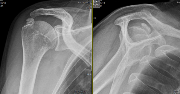
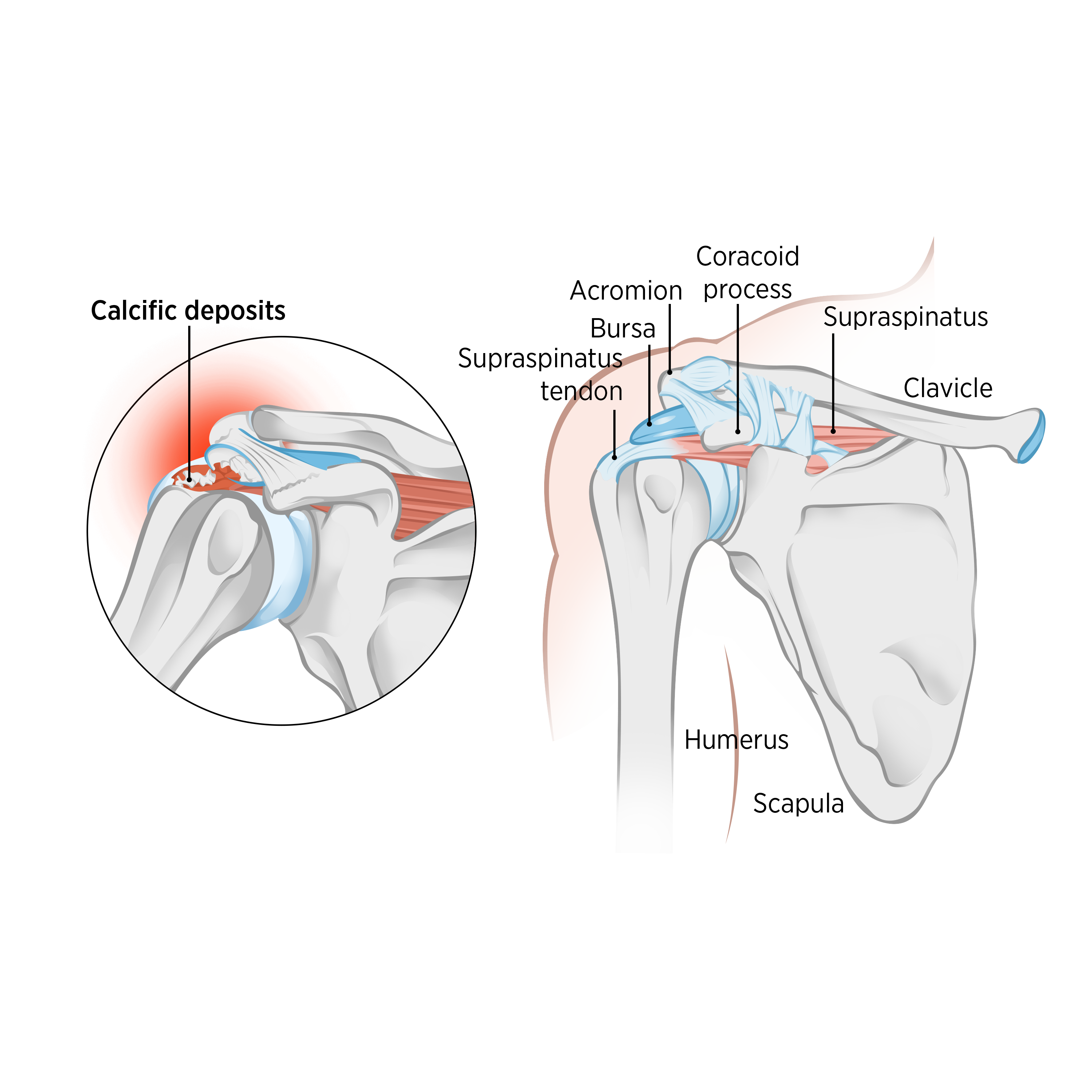
Calcific tendonitis of the shoulder is an acute or chronic painful condition due to the presence of calcific deposits inside or around the tendons of the rotator cuff. It is an enthesopathy, a self-limiting disease characterized by the deposition of calcium phosphate crystals in or around the rotator cuff tendons (see figure 1)(1). This calcium deposit is a paste-like material in or around the tendon and not a hard object that one would expect. An enthesopathy is a disease that affects places where tendons, ligaments, or muscles attach to bones.
Rotator cuff disease is a common cause of shoulder pain. Calcific tendonitis is one of many differential diagnoses of rotator cuff disease. In its acute presentation, patients typically respond to conservative treatment. While patients with chronic calcific tendonitis usually fail to respond to conservative treatment and often require surgery.
Among patients with calcific tendonitis, 2.7 – 20% are asymptomatic, and 35 – 45% of patients whose calcific deposits are discovered develop shoulder pain(1). It typically occurs between the ages of 30 and 50 and is rare in those older than 70. It is approximately twice as likely to occur in women as in men. It presents bilaterally in 10% of patients. Diabetes and gout are risk factors for calcific tendonitis. However, a comprehensive link is missing. Furthermore, there is no relation to injury, physical activity, diet, or osteoporosis.
Calcific Tendonitis Classification
There are several classifications of calcific tendonitis (see table 1). The classification used varies between practitioners and can be associated with the duration of clinical symptoms, degree of calcium invasion, association with or without endocrine disease, size, and morphology of the calcium deposit. For example, Neer’s classification divides pain and calcium deposit progress.
Table 1: Classification of calcific tendonitis(2-4).
| Classification | |
| Duration of Clinical Symptoms |
• Acute (with two weeks)
• Subacute (three to eight weeks)
• Chronic (more than eight weeks)
|
| Degree of Invasion | • Localized • Diffused – typically more painful and persists for longer |
| Association with Endocrine Disease | • Idiopathic calcific tendonitis (type 1) – not accompanied by endocrine disease. • Secondary calcific tendonitis (type 2) – accompanied by endocrine disease. Typically, they do not respond to conservative treatment. |
| Size of the Calcific Deposit | • Small - less than 0.5cm • Medium – 0.5 - 1.5cm • Large – greater than 1.5cm |
| Morphology of Calcium Deposit in Radiology | • Type 1 – fluffy shape with ill-defined margin (typically seen in acute phase – associated with pain) • Type 2 – dense with a well-defined margin (chronic phase with little pain). |
| Neer’s classification of pain and calcium deposit | • Type 1: Pain caused by chemical irritation because of the calcium deposit. • Type 2: Pain caused by increased local pressure in the tissue as it swells. • Type 3: Impingement-type pain due to bursal thickening and irritation by prominent calcium deposits. • Type 4: Pain caused by chronic stiffening of the glenohumeral joint. |
Clinical Presentation
Calcific Tendonitis Diagnosis
Treatment
Conservative management
The primary treatment of calcific tendonitis is conservative, with a 30 to 80% success rate(6,7). Nonsurgical treatment would include addressing the pain by taking oral painkillers and non-steroidal anti-inflammatory medication. While taking the medication, physiotherapy treatment can begin. Physiotherapy aims to create "space" in the glenohumeral joint. The calcium deposit takes up space either in or around the tendon. Manual therapy techniques assist in mobilizing the joint and soft tissue structures. Clinicians can prescribe gentle glenohumeral capsule, and rotator cuff muscle stretches to create space for the tendon to move within the subacromial space. Patients can start active assisted stretches in their home exercise program. The intention is to improve and maintain the range of motion and maximize functional levels.
Subacromial steroid injection is performed by specialists when pain levels are severe. Another treatment done by orthopedic surgeons is needle aspiration and irrigation(7). Extra-corporeal shockwave therapy is a more recent treatment choice(7). This therapy utilizes acoustic waves to induce fragmentation of the crystals. Furthermore, clinicians may use ultrasound-guided barbotage, where under ultrasound guidance, they inject the calcific deposit with a saline solution and then remove the calcium into a syringe. The area is then repeatedly washed out(6).
Surgical management
Clinicians may recommend surgery when conservative options fail to manage pain intensity(7). Surgery intends to reduce the effects of impingement and is typically arthroscopic, where surgeons make two or three mini-incisions around the shoulder joint. Through these key-hole incisions, they insert a camera and other surgical instruments to remove the calcium from the tendon under magnified vision. In its acute form, the calcium is paste-like, and surgeons can easily remove it with a fine needle. However, in its chronic presentation, the calcium tends to be more "stuck" and is not removed with as much ease. Depending on the size and location of the calcium deposit, the tendon is sometimes damaged when removing the deposit, requiring a complete surgical rotator cuff repair.
Post-surgery, there should be a dramatic relief of pain. Due to the phases of healing, a typical recovery in regaining range of motion and muscle power should be three to five months. This is influenced by how much of the calcific deposit occupied the tendon and thus affected when removing the calcium deposit.
The first phase of post-operative physiotherapy is to reduce the pain by managing swelling and inflammation around the glenohumeral joint. Electrotherapy, cryotherapy (ice application), and manual therapy are key modalities in the early healing phase. Once the pain and inflammation have settled, the next goal is to regain full shoulder range of motion. The physiotherapist should include accessory and physiological mobilization of the scapula and glenohumeral joints. Clinicians can include passive and active stretches to improve ROM. A home exercise program should include applying ice, gentle stretches, and introducing stability and strength exercises as the patient progresses through the tendon healing phases.
Conclusion
Calcific tendonitis is not a diagnosis that anyone would choose. However, several successful conservative and surgical solutions are available. Physiotherapy is a key part of either journey. The patient needs to wear their "patience cap" as there are no shortcuts to manage this complex diagnosis. Clinicians need to prioritize pain management to reduce the disuse consequences. Furthermore, they should manage the patient’s expectations and provide clear advice on recovery timelines and treatment options.
References
- J Shoulder Elb 2020 Dec; 23 (4): 210 – 216
- Clin Orthop. 1961; 20: 61 – 72
- R I Med J. 1953; 36: 512 – 516
- Neer CS II, editor. Shoulder Reconstruction. PA: WB Saunders; 1990. 421 – 485
- Br J Sports Med 2008. 42: 628 - 635
- Acta Biomed 2018; 89, 1: 186-196
- J Orthop Traumatol. 2016 Mar; 17(1): 7–14
Newsletter Sign Up
Subscriber Testimonials
Dr. Alexandra Fandetti-Robin, Back & Body Chiropractic
Elspeth Cowell MSCh DpodM SRCh HCPC reg
William Hunter, Nuffield Health
Newsletter Sign Up
Coaches Testimonials
Dr. Alexandra Fandetti-Robin, Back & Body Chiropractic
Elspeth Cowell MSCh DpodM SRCh HCPC reg
William Hunter, Nuffield Health
Be at the leading edge of sports injury management
Our international team of qualified experts (see above) spend hours poring over scores of technical journals and medical papers that even the most interested professionals don't have time to read.
For 17 years, we've helped hard-working physiotherapists and sports professionals like you, overwhelmed by the vast amount of new research, bring science to their treatment. Sports Injury Bulletin is the ideal resource for practitioners too busy to cull through all the monthly journals to find meaningful and applicable studies.
*includes 3 coaching manuals
Get Inspired
All the latest techniques and approaches
Sports Injury Bulletin brings together a worldwide panel of experts – including physiotherapists, doctors, researchers and sports scientists. Together we deliver everything you need to help your clients avoid – or recover as quickly as possible from – injuries.
We strip away the scientific jargon and deliver you easy-to-follow training exercises, nutrition tips, psychological strategies and recovery programmes and exercises in plain English.
