You are viewing 1 of your 1 free articles
Radial Tunnel Syndrome: A Rare Cause of Lateral Elbow Pain
Lateral elbow pain is common in athletes. The first suspicion is always lateral elbow tendinopathy, which is statically accurate. Evan Schuman explains what could be happening when athletes don’t recover as expected. Perhaps something has been missed.
Tennis - French Open - Roland Garros - Kazakhstan’s Elena Rybakina in action during her first-round match against Belgium’s Greet Minnen REUTERS/Gonzalo Fuentes
When typical management strategies fail, lateral elbow pain can create a conundrum for clinicians. Lateral elbow tendinopathy is the most common pathology and responds well to appropriate de-loading and gradual loading. However, what happens when things don’t go to plan? Radial tunnel syndrome (RTS) presents very similarly to lateral elbow tendinopathy and presents in 5% of patients diagnosed lateral elbow pain(1-3). Radial tunnel syndrome is a true dynamic entrapment neuropathy that causes lateral elbow and proximal dorsolateral forearm without significant motor fall-out(1-4).
Epidemiology
Radial tunnel syndrome is the most common cause of radial nerve branch entrapment, accounting for 1-2% of peripheral nerve entrapment in the upper limb. Moreover, it has a rare annual incidence of three in 100,000 cases. With such scarcity, one must wonder how many cases are misdiagnosed. Individuals with RTS often perform repetitive pronation and supination forearm movements of forearm pronation and supination. For example, manual laborers, machine workers, musicians, racquet sports, and heavy lifting(5). If these individuals present with lateral elbow pain, they most likely have lateral elbow tendinopathy, but clinicians must consider what else could be causing lateral forearm pain.
Anatomy
The Radial Nerve Pathway
The radial nerve is an extension from the posterior chord of the brachial plexus. It consists of nerve fibers from spinal nerve roots C5-T1. It arises in the axilla, where it separates from the posterior cord, anterior to the subscapularis, and travels along the posterior aspect of the upper arm(6,7). Then, it continues along the radial groove and supplies sensory and motor branches along its path(7).
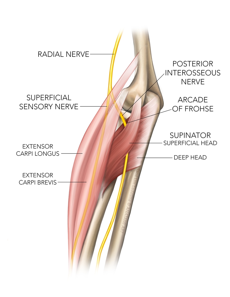
Approximately three centimeters distal to the lateral epicondyle, the radial nerve divides into two branches – the superficial branch of the radial nerve (SBRN) and the posterior interosseus nerve (PIN)(7). The SBRN is a cutaneous sensory nerve that innervates the dorsal skin on the lateral aspect of the hand and the proximal dorsal surfaces of the thumb, index, and lateral half of the third finger(8). The PIN pierces between the two heads of the supinator muscle. It provides motor innervation to the common and deep extensors extensor of the forearm and sensory innervation to the dorsal wrist capsule via unmyelinated (group IV) afferent fibers. These fibers are associated with nociception, which may be why patients with PIN entrapment may present with radiating pain(3). Notably, the PIN has no cutaneous innervation.
Radial Tunnel
The radial tunnel consists of four boundaries and extends from the head of the radius, anterior to the radiocapitellar joint, to the distal border of the supinator muscle, measuring approximately five centimeters in length (see table and figure 1).
Table 1: Radial tunnel boundaries(4,10)
| Boundary Anatomy | |
| Lateral | Mobile wad of Brachioradialis, Extensor Carpi Radialis Brevis, Extensor Carpi Radialis Longus |
| Medial | Biceps tendon and Brachialis |
| Floor | Capsule of the Radiocapitellar Joint |
| Roof | Supinator and Brachioradialis |
There are five potential irritation sites where the PIN may be compressed(11).
- Arcade of Froshe/Supinator Arch – The most common site of compression and the connection between the superficial and deep heads of the supinator muscle(12).
- Fibrous tissue anterior to the radiocapitellar joint.
- Leash of Henry – Recurrent radial vessels(13).
- The medial edge of the extensor carpi radialis brevis (ECRB).
- Distal edge of the supinator.
Along with the typically mechanical causes, additional aetiologies may result in nerve entrapment(4,7). These include tumors, foreign bodies, repetitive trauma, iatrogenic injections, and elbow arthritis.
Biomechanical Musculoskeletal Considerations
A biomechanical study using cadavers in Australia measured the pressure within the radial tunnel utilizing a balloon catheter during wrist flexion and forearm pronation. The study also assessed the forces exerted on the lateral epicondyle using a 1kg weight and transducer(10). The study results demonstrate that combined forearm pronation and wrist flexion cause a huge increase in pressure within the radial tunnel. Furthermore, under load, the supinator muscle significantly increases the force exerted on the common extensor tendon origin. Releasing it substantially reduces the radial tunnel pressure and forces at the lateral epicondyle. Finally, the researchers found that releasing the ECRB reduced the pressure within the radial tunnel.
Clinical Presentation
Athletes with PIN entrapment will present with localized achy pain approximately five centimeters distal to the lateral epicondyle. Furthermore, they may experience nerve-related night pain, which may interfere with sleep. Their symptoms are aggravated with combined forearm pronation and wrist flexion as these movements compress the PIN. Clinicians must assess grip strength as weakness may be present but is often from pain and not nerve involvement. Finally, they may have been unresponsive to treatment for lateral elbow tendinopathy(1,4).
Assessment
Radial tunnel syndrome is regarded as a diagnosis of exclusion, so clinicians must subjectively and objectively differentiate this condition. However, the presentation has similarities and differences (see table 2). A holistic assessment is critical, and clinicians must also consider cervical radiculopathy(1,4).
Table 2: Similarities and differences between RTS and lateral elbow tendinopathy
| Variable | RTS | Lateral elbow tendinopathy |
| Location | 4-5cm distal to the lateral epicondyle | Focal tenderness at the lateral epicondyle |
| Pain behaviour | No changes with a warm-up | No changes with a warm-up |
| Night pain | It may be present due to nerve involvement. | Absent |
| Test | ||
| Combined forearm pronation and wrist flexion | Produces pain | No pain |
| Mill’s Test | May reproduce symptoms | Produces pain |
| Resisted supination | Produces pain | No pain |
| Upper limb tension test – Radial nerve bias | Upper limb tension test – Radial nerve bias | It may reproduce symptoms due to forearm and wrist movement. Sensitization must be used to differentiate nerve-related symptoms from stretching of the common extensor tendons |
| Maudley’s test | Produces pain | Produces pain |
| Cozen’s test | It is unlikely to produce pain | Produces pain |
| Palpation | Pain 4-5cm distal to the lateral epicondyle | Pain more localized to the lateral epicondyle |
| Counterforce brace | Unlikely to provide relief | Some pain relief |
Imaging and Diagnostic Investigations
Clinicians need to know which studies or imaging will aid in diagnosing the suspected pathology, as this will inform the referral pathway.
Electrodiagnostic Studies
Electromyographic studies are inconclusive as the PIN carries fibers responsible for the conduction of pain and temperature changes. In RTS, the large, myelinated fibers are typically unaffected; therefore, the EMG studies are normal(7).
Diagnostic Injection
Clinicians can use a local anesthetic to confirm the diagnosis of RTS. An injection into the radial tunnel will not reduce symptoms of lateral elbow tendinopathy and vice versa(4).
Magnetic Resonance Imaging
Clinicians can use MRI to identify musculature atrophy caused by PIN entrapment and other causes of entrapment – tumors, ganglia, and radiocapitellar joint synovitis(4,7).
Management
Conservative management yields poor outcomes(14). Therefore, athletes typically require surgical decompression, which has a success rate of 67%-92%. There is a lack of literature on how to treat this condition best conservatively. Therefore, clinicians base their management plan on anatomical and physiological findings associated with RTS and applicable principles of managing other nerve-related pathologies(15). Where there is a high index of suspicion of RTS, clinicians should refer athletes early to an upper limb orthopedic surgeon.
Treatment goals
• Reduce pain.
• Restore full range of motion at the elbow and wrist.
• Restore muscle length.
• Restore neural mobility.
• Strengthening of the kinetic chain.
• Pain-free exercise.
• Prevent reoccurrence.
• Perform ADLs and sporting activities optimally.
Pain Reduction
If athletes decide against surgical intervention, clinicians should refer them to a physician to assist with pain management to facilitate function. This may be in the form of a cortisone injection or anti-inflammatories(16). Physicians may use ultrasound-guided corticosteroid injections to hydro dissect around the PIN(7). Splinting in positions of elbow flexion, forearm supination, and wrist extension may assist, but impracticality may inhibit adherence. A wrist splint to position the wrist in extension and to instruct patients to avoid positions of forearm pronation and elbow extension may be more practical(15). Furthermore, dry-needling of the supinator may assist(17).
Mobility
Clinicians must address neural mobility through radial nerve glides(15). However, they should carefully regulate the volume and intensity to avoid symptom flare-ups. Furthermore, they must avoid pain-provoking positions until the later rehabilitation stages. Athletes may present with stiffness at the wrist and elbow joints and muscles, and clinicians must restore mobility through joint mobilization and myofascial release techniques. Athletes can maintain their mobility with active range of motion exercises.
Strengthening
As the pain subsides, clinicians must prescribe strengthening exercises targeting the entire kinetic chain to improve the muscular capacity to tolerate load when returning to sports(15). These strengthening exercises need to be done in progression and pain-free positions. When loading the forearm muscles, starting with low sets, reps, and weight is advisable. Starting with isometrics and progressing to eccentric and concentric exercises may also be beneficial.
Ergonomics(15)
Clinicians must identify and address workplace and training contributions to pain during the first consultation. They must empower athletes to manage their condition as best as possible, take note of certain triggers, and avoid or manage them appropriately going forward.
Conclusion
Sports- and occupational-related lateral elbow pain is commonly seen in the clinical setting. Clinicians are pivotal in determining an accurate diagnosis and management plan for these patients. It is, therefore, essential to have a thorough understanding of the anatomy of the lateral elbow/forearm and the differential diagnoses in this region. To obtain an accurate diagnosis, clinicians must conduct a detailed subjective and objective evaluation, paying close attention to the similarities and differences between RTS and other pathologies in this region. When there is a high index of suspicion of RTS, it is essential to refer early on to achieve better patient outcomes sooner.
References
- J Ortho Sports Phys Ther.1991;14(1): 14-17
- J Plastic, Reconstr Aesthet Surg.2008; 61(9):1095-1099
- Arch Bone Jt Surg.2015;3(3):156–162
- J Hand Surg Eur Vol. 2020;45(8):882-889
- J Hand (N Y). 2023;18(1):146-153
- Anesth Pain Med. 2021; 14(11):(1)
- Curr Rev Musculoskelet Med. 2021 Jun;14(3):205-213
- Anatomy, Shoulder and Upper Limb, Radial Nerve. 2023 StatPearls
- J Hand Surg Am. 1979;4(1):52-59
- J Hand Surg Br. 2004 Oct;29(5):461-4
- J Hand Surg Am. 2023;48(11):1-7
- Surg Radiol Anat. 2005 Aug;27(3):171-175
- Neurosurg Rev. 2023 Feb 13;46(1):53
- J Hand Surg Am. 1983 Jul;8(4):414-420
- J Hand Ther. 2006;19(2):186-91
- Hand (N Y). 2019 Nov;14(6):741-745
- Physiother Theory Pract. 2019 Apr;35(4):373-382
Newsletter Sign Up
Subscriber Testimonials
Dr. Alexandra Fandetti-Robin, Back & Body Chiropractic
Elspeth Cowell MSCh DpodM SRCh HCPC reg
William Hunter, Nuffield Health
Newsletter Sign Up
Coaches Testimonials
Dr. Alexandra Fandetti-Robin, Back & Body Chiropractic
Elspeth Cowell MSCh DpodM SRCh HCPC reg
William Hunter, Nuffield Health
Be at the leading edge of sports injury management
Our international team of qualified experts (see above) spend hours poring over scores of technical journals and medical papers that even the most interested professionals don't have time to read.
For 17 years, we've helped hard-working physiotherapists and sports professionals like you, overwhelmed by the vast amount of new research, bring science to their treatment. Sports Injury Bulletin is the ideal resource for practitioners too busy to cull through all the monthly journals to find meaningful and applicable studies.
*includes 3 coaching manuals
Get Inspired
All the latest techniques and approaches
Sports Injury Bulletin brings together a worldwide panel of experts – including physiotherapists, doctors, researchers and sports scientists. Together we deliver everything you need to help your clients avoid – or recover as quickly as possible from – injuries.
We strip away the scientific jargon and deliver you easy-to-follow training exercises, nutrition tips, psychological strategies and recovery programmes and exercises in plain English.


