You are viewing 1 of your 1 free articles
Cover me! Understanding dermatology in Sports and Exercise Medicine – Part V
Wrapping up the series on dermatology related to sports medicine, Nella Grilo delves into the most lethal form of skin cancer.
Athletics—European Athletics Championships—Britain’s Katarina Johnson-Thompson shaded herself from the sun with an umbrella during the high jump in the women’s heptathlon. REUTERS/Aleksandra Szmigiel
Melanoma is skin cancer arising from melanocytes, and it is responsible for the majority of skin cancer-related deaths. Its aggressive nature and potential to metastasize demand early recognition and aggressive intervention. Prolonged and excessive exposure to ultraviolet (UV) radiation from the sun puts athletes at high risk for developing melanoma as they often spend extended periods in the radiant embrace of the sun during their training and competition.
Etiology
Melanoma primarily arises from the cumulative effects of exposure to UV radiation from the sun or tanning beds, which damages the DNA within skin cells. However, the disease’s complexity extends beyond UV exposure, with other contributing factors such as having fair skin, a history of severe sunburns -especially during childhood and adolescence - a family history of melanoma, the presence of numerous moles, a weakened immune system, and hormonal changes like pregnancy(1).
Types of Melanoma
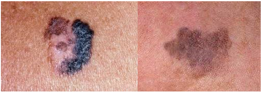
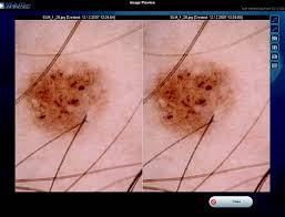
- Superficial Spreading Melanoma (SSM): This is the most common type of melanoma, accounting for approximately 70% of cases. It typically starts as a flat or slightly raised lesion with irregular borders. Over time, it may grow horizontally before penetrating deeper into the skin (see figure 1).
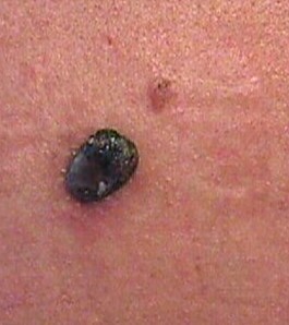
2.Nodular Melanoma (NM): This is a particularly aggressive variant, representing approximately 15-30% of cases. It typically presents as a dome-shaped nodule, often dark in color, and is prone to bleeding and ulceration (see figure 2). Unlike other forms, NM usually lacks the typical signs of asymmetry, irregular borders, and color variation. It grows quickly and is frequently diagnosed later, contributing to a less favorable prognosis.
3.Lentigo Maligna Melanoma (LMM): This type of melanoma usually affects older individuals with a history of extensive sun exposure. It typically develops slowly and initially appears as a flat, tan, or brown patch on the skin. Over time, it may become darker or develop irregular borders. It has a better prognosis than other subtypes when detected and treated early.
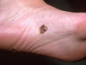
4. Acral Lentiginous Melanoma (ALM): This subtype primarily affects the palms of the hands, soles of the feet, or under the nails (see figure 3). It is more common in people with darker skin tones and is not strongly associated with UV exposure. It often starts as a dark spot or discoloration and may be mistaken for other non-cancerous conditions. Due to its location and diagnostic challenges, it is typically diagnosed at a more advanced stage, contributing to a less favorable prognosis.
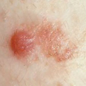
5. Amelanotic Melanoma: Amelanotic melanoma is a subtype of melanoma that lacks the typical dark coloration due to a lack of pigment production. It can appear as a flesh-colored or pinkish bump or lesion, making it easily mistaken for other benign skin conditions. Due to the difficulty in diagnosis, amelanotic melanoma is often detected at later stages, impacting prognosis.
6. Mucosal Melanoma: This can occur on any mucous membrane, such as those lining the mouth, nasal passages, gastrointestinal tract, and genitalia, and can appear as pigmented or non-pigmented lesions. Due to their hidden location and diverse appearance, they tend to be diagnosed at later stages and thus have a poor prognosis.
7. Ocular melanoma: Ocular melanoma is the second most common type after cutaneous and the most common primary intraocular malignant tumor in adults.
8.Desmoplastic Melanoma: This rare subtype tends to occur in sun-exposed areas, such as the head and neck. A fibrous or scar-like appearance characterizes it and can be challenging to diagnose. Desmoplastic melanoma has a higher tendency to spread along nerve pathways(2).
Detection and Diagnosis
Melanoma can appear as a new mole or develop within an existing mole. Recognizing the warning signs and seeking medical attention if any lesion exhibits the "ABCDE" characteristics: asymmetrical shape, irregular borders, color variation, diameter larger than six millimeters, and evolving characteristics. Other symptoms may include itching, bleeding, or a sore that does not heal. Not all melanomas exhibit these symptoms, so a healthcare professional should promptly examine any suspicious changes in the skin. Familiarity with these subtypes is crucial for dermatologists and healthcare professionals to ensure early and accurate diagnosis and to tailor treatment strategies accordingly.
Early detection is vital in pursuing better melanoma outcomes. Dermoscopy, a non-invasive technique, enhances diagnostic accuracy by magnifying skin lesions and recording images for follow-up comparison to detect subtle changes.
Practitioners should refer athletes to medical professionals who can remove any suspicious lesions. These are then biopsied or completely excised whenever possible. The primary care practitioner can perform this procedure easily, and there should be no delay in diagnosis while awaiting referral to a dermatologist if the lesion is deemed suspicious. The responsibility of diagnosis rests with the histopathologist, who is tasked with determining whether the lesion is atypical or overtly malignant and assessing for any invasion present and the depth of invasion.
Classification
Historically, clinicians used a Breslow depth and Clarke level when diagnosing invasive melanomas. The Breslow Depth is a helpful measure of how far melanoma has invaded the body. Knowing the depth of melanoma is useful because it is important when considering future treatment. However, the American Joint Committee on Cancer (AJCC) staging system has replaced the Breslow Depth.
The Clark Level is a staging system that describes the depth of melanoma as it grows in the skin. However, the current staging system adopted by the AJCC no longer considers the Clark Level. The Clark level is less prognostic and more subjective than other alternatives. The AJCC system assigns a stage based on tumor, node, and metastasis (TNM) (see table 1). The goal is that melanomas of the same stage will have similar characteristics, treatment options, and outcomes(3). There are various clinical stages, according to AJCC 8th edition. They range from Stage 0 to Stage IV.
Table 1: T classification of primary tumor for melanoma(4).
| T Category | Tumor Thickness | Additional Prognostic Parameters |
| Tis | Melanoma in situ, no tumor invasion | |
| Tx | No information | Tumor thickness cannot be determined |
| T1 | ≤1.0 mm | a: <0.8 mm, no ulceration b: <0.8 mm with ulceration or 08-1.0 mm with or without ulceration |
| T2 | >1.0-2.0 mm | a: No ulceration b: Ulceration |
| T3 | >2.0-4.0 mm | a: No ulceration b: Ulceration |
| T4 | >4.0 mm | a: No ulceration b: Ulceration |
Treatment
Treatment for melanoma depends on various factors, including the stage of the cancer, the location and size of the tumor, and the individual’s overall health. Surgery to remove the tumor is the primary treatment for all melanoma stages. The ESMO Clinical Practice Guidelines of 2019 are as follows:
- In situ: 0.5cm
- <2mm tumor thickness: 1cm
- >2mm tumor thickness: 2cm
A sentinel lymph node biopsy is almost always indicated in lesions with a depth of ≥1.0 mm. If the sentinel node is positive, further staging becomes necessary, and these patients should be referred to an oncologist. Oncologists may request additional diagnostic tests, including blood tests, CT scans, and PET scans, to assess the potential metastasis of the tumor.
Previous therapies for systemic metastatic disease have yielded low response rates. However, recent advancements in drug development show significant promise for patients with systemic melanoma metastases. Immunotherapy drugs known as checkpoint inhibitors are the primary treatment. These drugs can shrink tumors over extended periods in some individuals. While combinations of checkpoint inhibitors appear to be more effective, they also carry a higher risk of severe side effects.
Approximately half of all melanomas exhibit BRAF gene changes. These melanomas often respond well to targeted therapy drugs, typically involving a combination of BRAF and MEK inhibitors. However, clinicians usually attempt to use immune checkpoint inhibitors as the initial treatment, as they provide longer-lasting benefits. Alternatively, they can consider a combination of targeted drugs alongside an immune checkpoint inhibitor. However, these drugs’ intricate mechanisms of action and the selection of appropriate combinations are complex(5).
Prognosis
Lesions in their early stages, with minimal or no invasion, generally have an excellent prognosis. However, the prognosis rapidly deteriorates as the depth of invasion increases (see table 2), emphasizing the importance of early detection.
Table 3: Melanoma survival rate
| Stage | Survival Rate |
| 0 | 99.9% 5-year survival |
| I/II | 89-95% 5-year survival |
| II | 45-79% 5-year survival |
| III | 24-70% 5-year survival |
| IV | 7-19% 5-year survival |
Due to the increased risk of developing a new melanoma compared to the general population, athletes with a history of melanoma should seek regular follow-ups. Stage 0 in situ melanoma necessitates at least annual skin examinations for life and monthly self-examinations without routine blood tests or imaging.
Patients with stage IA-IIA melanoma should have history and physical examinations every 6-12 months for five years, then annually, along with monthly self-examinations. Routine blood tests and imaging are not recommended unless specific signs or symptoms arise.
For stage IB-IV melanoma patients with no evidence of disease, follow-up includes history and physical examinations every 3-6 months for two years, then every 3-12 months for another two years, and annually after that, with imaging as indicated. Routine blood tests are not recommended, and routine imaging is discouraged after five years unless mandated by clinical trial participation(7).
Conclusion
Melanoma is a serious concern for athletes who spend significant time in the sun. Despite outdoor activities’ allure, athletes must prioritize sun safety measures to reduce their risk of developing melanoma. Regular skin checks, early detection, and prompt medical attention for suspicious lesions are imperative. Additionally, understanding the importance of follow-up appointments and adhering to recommended surveillance schedules can significantly impact long-term outcomes. By adopting sun-safe behaviors and remaining vigilant about skin health, athletes can continue to pursue their passion for outdoor activities while minimizing the risk of melanoma. Remember, protecting the skin today ensures a healthier tomorrow.
References
1. Barnhill RL, Mihm MC, et al. “Malignant melanoma.” In: Nouri K, et al. Skin Cancer. McGraw Hill Medical, China, 2008: 140-167
2. Cancer. 1999 Jul 15;86(2):288-99
3. www.molemaxsystems.com/wp-content/uploads/2021/12/MoleMax-2022- Current-Range-Brochure-V3.pdf
4. [Guideline] National Comprehensive Cancer Network. NCCN Clinical Practice Guidelines in Oncology: Melanoma: Cutaneous. Available at www.nccn.org/professionals/physician_gls/pdf/cutaneous_melanoma.pdf. Version 2.2023 — March 10, 2023; Accessed: October 26, 2023
5. Cancer Biol Ther. 2019; 20(11): 1366–1379
6. Annals of Oncology. 2020: 31(11):1435–1448
7. Melanoma: Staging and Follow-Up July 2021 Dermatology Practical & Conceptual 11(Suppl 1):2021162
Further reading
Newsletter Sign Up
Subscriber Testimonials
Dr. Alexandra Fandetti-Robin, Back & Body Chiropractic
Elspeth Cowell MSCh DpodM SRCh HCPC reg
William Hunter, Nuffield Health
Newsletter Sign Up
Coaches Testimonials
Dr. Alexandra Fandetti-Robin, Back & Body Chiropractic
Elspeth Cowell MSCh DpodM SRCh HCPC reg
William Hunter, Nuffield Health
Be at the leading edge of sports injury management
Our international team of qualified experts (see above) spend hours poring over scores of technical journals and medical papers that even the most interested professionals don't have time to read.
For 17 years, we've helped hard-working physiotherapists and sports professionals like you, overwhelmed by the vast amount of new research, bring science to their treatment. Sports Injury Bulletin is the ideal resource for practitioners too busy to cull through all the monthly journals to find meaningful and applicable studies.
*includes 3 coaching manuals
Get Inspired
All the latest techniques and approaches
Sports Injury Bulletin brings together a worldwide panel of experts – including physiotherapists, doctors, researchers and sports scientists. Together we deliver everything you need to help your clients avoid – or recover as quickly as possible from – injuries.
We strip away the scientific jargon and deliver you easy-to-follow training exercises, nutrition tips, psychological strategies and recovery programmes and exercises in plain English.


