You are viewing 1 of your 1 free articles
Ligamentous Ankle Injuries: Clinical Clues
Ankle injuries account for up to 40% of all sports injuries and are often seen in the physiotherapy clinic. Many have long-term residual symptoms, and up to 20% develop chronic instability. Samantha Nupen explores good clinical practice to ensure a comprehensive assessment to guide management.
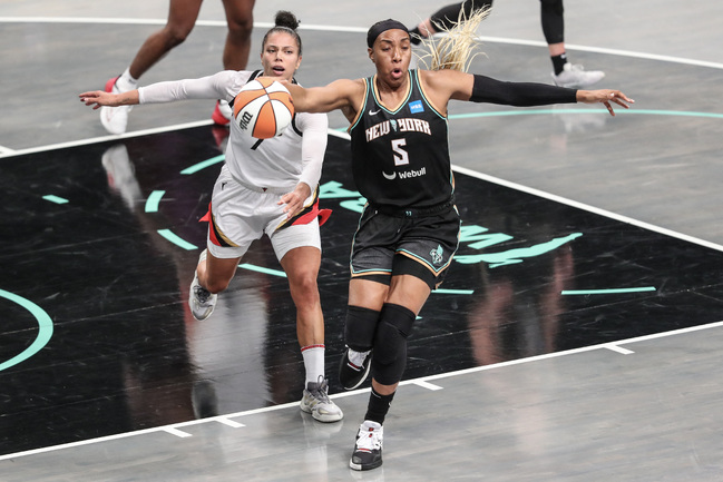
New York Liberty forward Kayla Thornton jumps in front of Las Vegas Aces forward Alysha Clark in the fourth quarter to chase down the ball during game three of the 2023 WNBA Finals at Barclays Center. Mandatory Credit: Wendell Cruz-USA TODAY Sports
Acute assessment
When a patient with an ankle injury hops or hobbles into the rooms, the first clinical decision to make is whether to send them for an x-ray. The severity of the swelling and bruising rarely corresponds to the severity of the injury. The Ottawa Ankle Rules (OAR) are reliable to help make this decision(1).
- As per the OAR, patients only require ankle and foot X-rays if they have one of the following criteria:
- Bony tenderness along distal 6 cm of the posterior edge of the fibula or tip of the lateral malleolus,
- Bony tenderness along distal 6 cm of the posterior edge of tibia/tip of medial malleolus,
- Bony tenderness at the base of the fifth metatarsal,
- Bony tenderness at the navicular,
- If they could not bear weight immediately after injury and for four steps during the initial evaluation.
Clinicians can use anteroposterior and lateral views to exclude lateral malleolus or distal fibula fractures, the most common ankle fracture.
Fractures of the base of the fifth metatarsal are commonly associated with severe ankle sprains. Avulsion fractures account for 93% of these, with Jones fractures less common but important not to miss because they take longer to heal due to poor circulation (see figure 1).
The other fracture associated with severe ankle sprains is a Maisonneuve fracture of the proximal fibula, especially if there is a rotational component in the mechanism of injury. This is a spiral fracture often associated with a syndesmosis injury. If this fracture is present, clinicians must assess for an associated fracture of the medial malleolus and possible complete rupture of the deltoid ligament complex.
Ankle Ligament Injuries
Lateral ligament complex
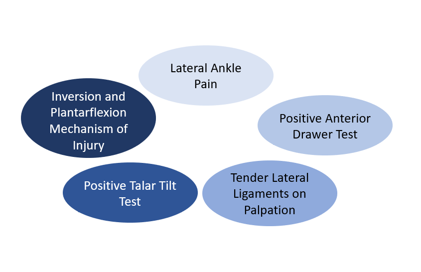
The most common ankle injury is a lateral ligament sprain. The mechanism of injury is typically combined plantarflexion and inversion, which places an excessive load on the lateral ligament complex. The anterior talofibular ligament (ATFL) resists torsion and inversion stress in plantarflexion and is the weakest and most commonly injured. The calcaneofibular ligament (CFL) is extra-articular and resists torsion and inversion in dorsiflexion. Finally, the posterior talofibular ligament (PTFL) resists posterior movement of the talus, is the strongest ligament, and is hurt the least. Injuries to the PTFL usually only occur in severe ankle sprains, which also involve the ATFL and CFL.
Lateral ankle sprains are graded in two ways: by the number of ligaments involved and by the severity of the injury. A grade three lateral ligament complex sprain involves all three lateral ligaments, but an athlete could sustain a grade three ligament sprain (complete rupture) to only the ATFL.
Clinical Assessment
The anterior drawer tests the ATFL and has a specificity of 80% five days post-injury(2). This test glides the calcaneus anteriorly while applying posterior counterpressure on the distal fibula and tibia in neutral plantarflexion. Pain and excessive gliding is a positive test with an increased glide of 4-5mm, indicating an ATFL tear.
The talar tilt/inversion stress test has a sensitivity of 52% and tests the CFL(3). With the ankle in neutral, the clinician inverts the heel, with the talus and calcaneus moving as one unit to avoid subtalar inversion. Pain over the CFL or a clunk is a positive test. An outward translation of >5o with a spongy or empty end feel indicates a complete rupture (see figure 2).
Medial/deltoid ligament complex
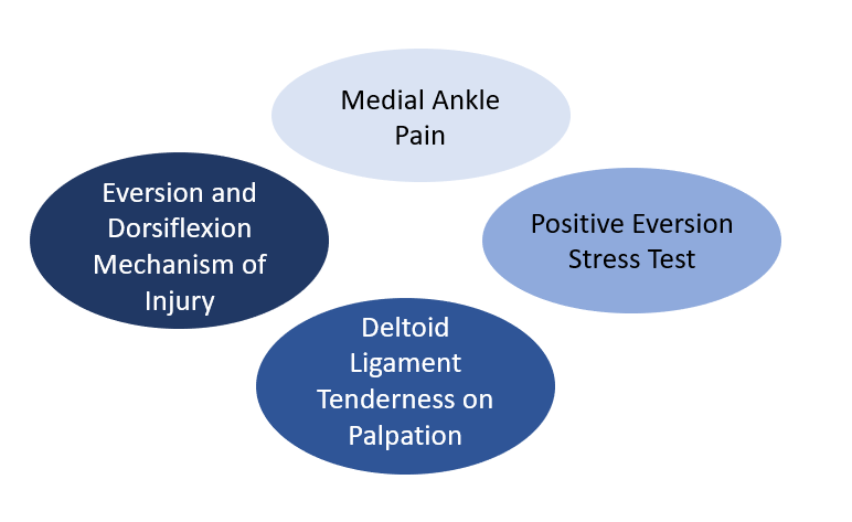
The deltoid ligament complex is stronger than the lateral ligaments and is only sprained in a severe injury. The deltoid ligament stabilizes the talotibial joint and transfers forces between the tibia and tarsus. It fixates the tibia above the talus and restricts the talus from shifting into a valgus position, translating antero-laterally, or rotating externally.
Clinical Assessment
The eversion stress test/eversion talar tilt test has a 96% sensitivity and 84% specificity five days post-injury for the deltoid ligament complex(4). Clinicians evert and abduct the heel while stabilizing the lower leg. The test is positive if there is pain and laxity on the medial side of the ankle. A spongy or empty end feel indicates a complete rupture.
Syndesmosis Injury(5)
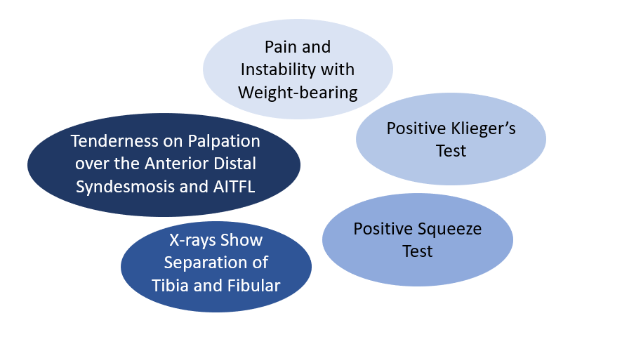
A syndesmotic, or ‘high’ ankle sprain involves the ligaments binding the distal tibia and fibula at the distal tibiofibular joint and may lead to ankle instability. Injuries can occur with any ankle motion, but the most common motions are extreme external rotation or dorsiflexion of the talus. The talar dome is broader anteriorly than posteriorly, and these movements force the ankle mortise apart, separating the medial and lateral malleoli. The sufficient distraction can cause strain or rupture to the anterior inferior tibiofibular ligament (AITFL), superficial posterior inferior tibiofibular ligament (PITFL), transverse tibiofibular ligament, and interosseous membrane (see figure 4). Injuries are also commonly present with fractures of either malleolus or a proximal fibular spiral fracture (Maisonneuve fracture).
Clinical Assessment
Kleiger’s test/external rotation test has a sensitivity of 75% and assesses the anterior tibiofibular ligament and syndesmosis. The test is positive if there is pain over the inferior tibiofibular syndesmosis with foot external rotation while stabilizing the leg. Athletes may experience pain over the deltoid ligament, or it may radiate upwards into the syndesmosis.
The squeeze test/fibular compression test assesses the anterior tibiofibular ligament and syndesmosis. Clinicians compress the tibia and fibula around the calf midpoint, which will reproduce pain in the distal syndesmosis area if it is positive.
To immobilize with bracing or taping?(6-8)
The effects of ankle taping and bracing on proprioceptive input to the central nervous system, peroneal muscle activity, and deceleration of ankle motion may be as important as restricting ankle ROM following injury. Initially, clinicians can recommend bracing or taping to protect and provide an optimum environment for healing. As athletes progress through rehabilitation, they can use bracing or taping to prevent another ankle injury.
Bracing vs. taping
- Cost: A brace is a once-off cost, whereas tape costs are ongoing.
- Skin condition: Repeated taping may negatively affect skin condition.
- Sport-specific legislation and equipment limitations, e.g., wearing taping with tight footwear may be easier.
- Access to professionals to apply the tape effectively.
Clinical Management
Clinicians must guide their clinical management according to a thorough assessment. Although pain and swelling may be the acute signs following injury, a wide range of neuromuscular skeletal deficits will impact rehabilitation and return to sport. Clinicians must continue to assess and re-evaluate these deficits and adjust rehabilitation to meet the demands of the sport for the injured athlete. Below are critical areas of concern and their respective assessment tools to reassess adequate recovery.
- The knee-to-wall/standing lunge test assesses the passive ankle dorsiflexion range. Assessing and monitoring ankle dorsiflexion range of motion is essential, aiding in evaluating functional recovery and preventing long-term mobility limitations. It is important to compare the affected and unaffected limbs.
- Assessing soleus and gastrocnemius muscle length after an ankle injury is crucial to identify and address potential muscle tightness and imbalances that impact ankle stability, joint function, and the risk of future injuries.
- Maintaining balance through the single-leg stance (SLS) test is essential. While age-specific norms typically range between 45 to 60 seconds, it’s important to remember that superior balance control significantly enhances sports performance, making it a critical factor for overall athletic success.
- Proprioception plays a crucial role in balance control. Distal proprioception tests and contralateral joint matching tasks assess proprioception. Ankle proprioception correlates strongly with competitive sport level and is the most significant predictor of sports performance. Ankle proprioception provides essential information to adjust ankle positions and upper body movements to successfully perform the complex motor tasks required in elite sports(9,10).
- Calf muscle strength and endurance are essential for good performance in sports. Clinicians can use the heel rise test (HRT) to assess calf capacity. The median in healthy adults is 24 for males and 21 for females(11). However, clinicians must note the pre-injury physical condition of the athlete as some may need to surpass population norms due to participating in high-demand sports, e.g., long-distance runners.
- Foot posture, function, and biomechanics ensure a strong medial arch to create a robust springboard for jumping, running, and acceleration. The ability to absorb and transfer force through the foot and ankle is critical for performance and resilience against injury.
Researchers in the Netherlands reviewed the preventive effectiveness of neuromuscular training (NMT) in reducing ankle sprains. The analysis of 30 studies, including various types of interventions, found that NMT, particularly balance training, significantly reduces the occurrence of ankle sprains in both athletes with previous ankle injuries and those without. While the evidence for preventing first-time ankle sprains is inconclusive, both single-component and multicomponent NMT interventions appear effective, emphasizing the importance of selecting appropriate interventions based on the context(12).
Conclusion
Ankle ligament injuries are commonly seen in the physiotherapy clinic. The diagnosis and management are based on clear and thorough assessment and the many clinical clues provided by the patient. This will lead to sound clinical decision-making and a successful return to sport for the athlete.
References
- BMC Musculoskeletal Disorders. 2022; 23:885.
- J Bone Joint Surg Br 1996 Nov;78(6):958-62.
- Clinical Radiology. 1986;37(3):247-251.
- www.physio-pedia.com/Stress_tests_for_Ankle_ligaments
- radiopaedia.org/articles/distal-tibiofibular-syndesmosis-injury
- J Athl Train. 2002; 37(4):436–445.
- JISAKOS 2016; 1:304–310.
- PLOS ONE | DOI:10.1371/journal.pone.0124214, 2015
- Biomed Res Int. 2015; 842804.
- J Sport Health Sci. 2016; 5(1): 80–90.
- J Physio. 2017; 103 (4):446-452.
- JISAKOS 2016; 1:202–213.
Newsletter Sign Up
Subscriber Testimonials
Dr. Alexandra Fandetti-Robin, Back & Body Chiropractic
Elspeth Cowell MSCh DpodM SRCh HCPC reg
William Hunter, Nuffield Health
Newsletter Sign Up
Coaches Testimonials
Dr. Alexandra Fandetti-Robin, Back & Body Chiropractic
Elspeth Cowell MSCh DpodM SRCh HCPC reg
William Hunter, Nuffield Health
Be at the leading edge of sports injury management
Our international team of qualified experts (see above) spend hours poring over scores of technical journals and medical papers that even the most interested professionals don't have time to read.
For 17 years, we've helped hard-working physiotherapists and sports professionals like you, overwhelmed by the vast amount of new research, bring science to their treatment. Sports Injury Bulletin is the ideal resource for practitioners too busy to cull through all the monthly journals to find meaningful and applicable studies.
*includes 3 coaching manuals
Get Inspired
All the latest techniques and approaches
Sports Injury Bulletin brings together a worldwide panel of experts – including physiotherapists, doctors, researchers and sports scientists. Together we deliver everything you need to help your clients avoid – or recover as quickly as possible from – injuries.
We strip away the scientific jargon and deliver you easy-to-follow training exercises, nutrition tips, psychological strategies and recovery programmes and exercises in plain English.
