11 factors that differentiate sciatica from hamstring or other causes of posterior thigh pain
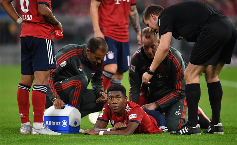
Posterior thigh pain provides a great challenge to sports physiotherapists around the world, with the most common cause being hamstring strains(1). However, there are a number of structures that cause pain both locally or referred. Identification of the pain source allows the practitioner to effectively manage the return to sport. Ultimately one must ascertain if the injury to the posterior thigh is an acute muscle injury or referred pain from another source. An understanding of the relevant anatomy of the posterior thigh, sacroiliac joint, and lumbar spine is therefore essential for effective diagnosis.
Anatomy
The hamstrings consist of the bicep femoris (BF), semimembranosus (SM) and semitendinosus (ST) (see figure 1). The ST and the BF unite to form a conjoined tendon before attaching to the posteromedial portion of the ischial tuberosity. The SM is considered to have a deeper attachment, originating just anterolaterally to the conjoined attachment on the ischial tuberosity. The adductor magnus also shares a common attachment to the medial border of the ischial tuberosity. It’s important to note that the proximal tendon has an intimate relationship with the inferior gluteal nerve and artery, the sciatic nerve and the adductor insertion. This can lead to proximal hamstring or adductor entities being accompanied by a neural irritation.Figure 1: Major muscles of the rear thigh

Subjective examination
The importance of a detailed history cannot be overstressed in the differential diagnosis of posterior thigh pain. A strain to the hamstring muscles occurs as a result of significant force. The individual will remember a specific incident such as sprinting or excessive eccentric force (eg the ‘Jackal’ position in rugby union with forced hip flexion whilst in relative knee extension). The mechanism of injury (MOI) is a key tool in the differential diagnosis of posterior thigh pain; if the athlete cannot provide an MOI and there are no local muscles signs, one should consider pain to be referred from another source.The number of potential causative structures can be significantly reduced via appropriate questioning and reasoning. The working diagnosis is further refined via appropriate structuring of the objective examination (see box 1).
Box 1: 11 Factors that differentiate the diagnosis
- Symptom onset– Was there an incident with no precipitating signs? (a) Yes – consider a strain, fascial or neural trauma. (b) No – consider overuse injuries, referred pain, other pathological entity.
- Location of symptoms– Are symptoms more proximal in nature with pain on direct palpation (a) Yes – consider all local structures. (b) No, distal symptoms increases the likelihood of a strain (especially in conjunction with a mechanism). However, referred pain and neural compromise will need to be cleared(2).
- Presence of neurological symptoms – Any sign of nerve involvement must lead to a thorough neurological and lumbar examination.
- Causative factors– Were there any pre-existing symptoms? A review of potential causative factors will lead the practitioner to eradicate or at the very least modify the provocative activity. For example, an ineffective Roman deadlift technique leading to proximal hamstring shear against the ischial tuberosity should be removed from the program initially until symptoms have resolved and correct technique can then be graded into the programming(3).
- Progress after injury– (a) Slow progress indicates a more severe injury. (b) Variable/erratic progress makes a strain improbable or indicates symptoms are being aggravated by activity. This requires a detailed review of current management and daily activities to identify the aggravating activities.
- Response to activity– Symptoms which increase with activity and are worse after, represent an inflammatory pathology. If an athlete starts training with minimal or no pain, that builds during but is not as severe on termination of training, neurological or vascular claudication must be considered(4,5).
- Recurrent problem– Does the athlete have a past medical history of posterior thigh injury? This requires extensive examination and a biomechanical review to ascertain potential movement patterns that could predispose an individual. An individual with recurrent hamstring strains requires a thorough review to address the root cause of the problem. Attending to the strain alone will not provide the athlete with the resilience to avoid re-injury.
- Level of activity– (a) Has a change in training coincided with the injury? (b) Is there an adequate base of training? A spike in loading is commonly the cause of injury - be that total volume (may indicate more overuse patterns such as proximal hamstring tendinopathy or gluteal tendinopathy) or sprint distance (likely to predispose the individual to strains)(6).
- Lifestyle/occupation– What other factors external to sport aggravate the condition? Prolonged sitting through extended periods of driving or desk work or repeated flexion movements to pick up young children can all exacerbate symptoms.
- Age and gender of the patient– Although not a key indicator, in the absence of an obvious mechanism, there are potential pathologies that a younger athlete will be at risk of, such as traction apophysitis. Female athletes may also have a greater risk of stress fractures due to the female athlete triad(7).
- Night pain– This could indicate a more sinister pathology, especially in the absence of a sports-related mechanism. As with all red flags, a review of the athlete with a doctor is essential who may then book further investigative scans or reviews(8).
Objective examination
The objective examination that follows the subjective history will further refine your working hypotheses. A pragmatic approach to assessment will ensure all potential pathologies have been considered. An ability to conduct a thorough objective examination is assumed; however, points that the author considers especially pertinent to each potential diagnosis are highlighted in table 1.| Possible differential diagnosis | Exclusion criteria |
|---|---|
| Lumbar spine facet arthropathyDisc degenerationRadiculopathy | No diffuse leg referral+ve hamstring load testsLumbar palpation (NAD) & -ve quadrant test |
| Hip joint ischiofemoral impingement+/- Quadratus femoris abnormalities | Femoral external rotation in hip neutral -veMRI -ve (No loss of space and QF normal)Flexion-adduction-internal rotation (FADDIR) -ve |
| SIJ somatic referral | Lasletts’s SIJ provocation tests -ve |
| Sciatic nerve compression | Sciatic tenderness at QF -veSlump test +ve hamstring but no change with sensitisers (Hip adduction/internal rotation)Modified slump (lx extension) Differential test for specific comparison to PHT*Coexisting pathology possible |
| Deep gluteal syndrome/ Piriformis syndrome | Sciatic nerve non-tender at piriformisNo further provocation with piriformis stretch/contraction or slump with Add/IRMRI imaging -ve |
| Gluteal tendinopathy | +ve Hamstring load tests-ve gluteal load testsMRI -ve |
| Ischiogluteal bursitis | Pain with stretch and localised palpationIrritable symptoms with sittingMRI and ultrasound -ve |
| Partial or complete tear of the gluteal or hamstring muscle/tendon | Gluteal and hamstring tests +ve but MRI and U/S findings -ve for muscle or tendon tear respectively |
| Posterior pubic or ischial ramus stress fracture | Tenderness over ischial ramusMRI imaging -veHigh suspicion in female triad athletes |
| Adductor magnus pathology, tear or tendinopathy | Adductor tests -ve,PSST adductor stretch and resist -veMRI -ve |
| Vascular endofibrosis | No immediate resolution of symptoms on cessation of provocative exercise.Bruit sign -veEchography or arteriography -ve |
Hamstring strains
Acute hamstring strains are often our first thought when a player grabs at their posterior thigh. Local pain with tenderness on direct palpation, loss of range and loss of muscle power can help confirm this diagnosis. The single leg bridge is a recognized quick and easy test to assess hamstring function. Tendon involvement will lengthen the time frame and likely alter the management plan, imaging will confirm this(10). Table 2 provides an easily digestible comparison between hamstring strain and tendinopathy to surrounding structures and referred pain.Avulsion of the hamstring tendon - although not common - may occur in the sporting population; an older runner with chronic proximal hamstring tendinopathy may rupture from its proximal bony attachment. In the younger population, (aged 14-18 years) avulsions of the ischial apophysis can occur. This will present as a high hamstring injury, and imaging will help clarify the extent(11). In adults, complete rupture is rare but may occur with sudden forced hip flexion and knee extension, such as in powerlifting and rugby(12,13).
| Clinical features | Acute hamstring strain(Type I or II) | Hamstring/Adductor/Gluteal Tendinopathy | Referred pain to the posterior thigh |
|---|---|---|---|
| Onset | Sudden | Gradual onset, progressive over time | May be sudden onset or gradual feeling of tightness |
| Pain | Moderate to severe | Low to moderate | Usually less severe, may feel like cramping or twinge |
| Ability to walk | Disabling - Difficulty walking, unable to run | Often able to walk pain free | Often able to walk / jog pain free |
| Stretch | Markedly reduced | Combined hip flexion and knee extension reduced range with possible symptom reproduction | Minimal reduction |
| Strength | Markedly reduced contraction with pain against resistance | May be reduced when assessed in hip flexion position | Full or near to full muscle strength against resistance |
| Local signs | Hematoma, bruising | None | None |
| Tenderness | Focal tenderness | Deep palpation of proximal or distal tendon reproduces symptoms | Variable tenderness, usually non-specific |
| Slump test | Negative | Occasionally positive due to proximity to neural tissue | Frequently positive |
| Trigger points | May have secondary gluteal trigger points | May have secondary gluteal and adductor trigger points | Gluteal or adductor magnus trigger points that reproduce hamstring pain on palpation or needling |
| Lumbar spine / SIJ signs | May have abnormal lumbar spine / SIJ signs | May have abnormal lumbar spine / SIJ signs | Frequently have abnormal lumbar spine / SIJ signs |
| Investigations | Abnormal ultrasound / MRI | Possibly abnormal ultrasound / MRI | Normal ultrasound / MRI |
Referred pain
In the absence of clear muscle injury signs and symptoms, referred pain must be considered. Posterior thigh pain could be caused by the lumbar spine, sacroiliac joint, hip joint, or neural or vascular compromise, and these must all be fully excluded. The slump test should be used to detect neural mechano-sensitivity. A positive slump test is strongly suggestive of referred pain. However, a negative slump doesn’t exclude this possibility(14). It is important to note that predisposing factors for hamstring injury may be found when the detailed proximal assessment has been completed.Trigger points are common sources of referred pain to the posterior thigh. The most common trigger points that refer pain to the hamstring are in the gluteus minimus, medius and piriformis(15). The classic presentation will be a feeling of tightness or cramping and the athlete may report that the hamstring feels like it is ‘on the edge of a strain’. Further examination may highlight restriction within the hamstring and the gluteals, but with direct referral into the hamstring on trigger point palpation.
Lumbar spine and other articular structures
The lumbar spine is a common source of posterior thigh pain. Pain may be referred from a number of structures such as discs, facet joints, muscles, ligaments, nerve roots(16). A detailed examination including a thorough neurological screen is vital, and imaging may help with this. However, caution is advised as pre-existing changes may be present. Nerve root compression will usually provide a much more definitive presentation. One must always consider red flags; cauda equina identification is vital to allow appropriate and timely management(17). Spondylolisthesis and spondylolysis have both been associated with hamstring pain and tightness and should be excluded in recurrent/recalcitrant presentations.The sacroiliac joint (SIJ) often refers pain to the buttocks and high hamstring region; 94% of SIJ dysfunction cases report buttock pain(18). SIJ provocation tests can be used to highlight the involvement of the SIJ(19). Anterior hip impingement can present with groin pain, lateral hip/thigh pain and buttock pain(20). A negative FAIR/FABER test is commonly used to rule out significant FAI and/or labral pathology(21).
Deep gluteal syndrome
Deep gluteal syndrome (DGS) is an umbrella term that covers a number of pathologies. Piriformis syndrome, compressing the sciatic nerve is the most common(22), but there can also be fibrous bands tethering the sciatic nerve, compression within the Gemelli–obturator internus complex, vascular pathologies or space-occupying lesions. Piriformis overactivity is a common response to weak gluteals; the need to assess the biomechanical control of athletes will allow for possible causative factors to present themselves. McCory and Bell highlight that it is also the hip external rotators that can compress the sciatic nerve(23). A combination of the seated pirformis stretch test and active piriformis test has high sensitivity and specificity for sciatic nerve entrapment(24).Vascular
Endofibrosis of the external artery can produce posterior thigh pain (although very rare compared to the more common presentation of lateral and anterior thigh pain). This can occur in cyclists or triathletes. Pain is claudicant in nature and progresses within 10-20mintues of exercise, but resolves immediately on termination of exercise.In summary
When assessing posterior thigh pain, the practitioner should take a stepwise approach, first forming a working diagnosis based on subjective findings, having excluded any red flags. A thorough objective assessment can then either rule in or rule out the working hypotheses. It is important we don’t allow our own confirmation bias to guide us to the most common pathology. We owe it to the athlete to ensure we have excluded as many potential causes prior to our definitive diagnosis. This allows implementation of the most appropriate evidence-based management plan and a prompt return to sport.References
- Br J Sports 2012; 46: 86-7.
- B Prac Res Clin Rheumatol 2007; 21 (2): 261-77.
- Br J Sports 2012; 46: 833-834.
- Sports Med 2002; 32: 371-91.
- Eur J Vasc Endo Surj 2003; 26 (6).
- Br J Sports Med 2016; 0: 1-9.
- Military Med 2011 176; 420-430.
- BMJ 2013; 347: f7095.
- Sports Physio 2017; 2: 20-24.
- Am J Sports Med 2012; 41 (1): 111-115.
- Clinical Sports Medicine 2012; 30: 600.
- Am J Sports Med 1998; 26 (6): 785-8.
- Knee Surg Sports Traumatol Arthrosc 2008; 16: 797-802.
- JCR 2008; 14 (2): 87-91.
- B J Sports Med 2005; 39 (2): 84-90.
- Spine 2002; 27 (22): 2538-2545.
- Br J Neurosurg; 2010; 24 (4): 383-6.
- Arch Phys Med Rehabil 2000; 81 (3): 334-8.
- Arthroscopy J 2005; 10 (3): 207 – 18.
- Clin Orth & Rel Res 2009; 467 (3): 638-644.
- Scand J Med Sci Sports 2017 27 (3): 342-350.
- Am J Sports Med 1998; 16 (5): 517-21.
- Sports Med 2002 32 (6) 371-91.
You need to be logged in to continue reading.
Please register for limited access or take a 30-day risk-free trial of Sports Injury Bulletin to experience the full benefits of a subscription. TAKE A RISK-FREE TRIAL
Newsletter Sign Up
Subscriber Testimonials
Dr. Alexandra Fandetti-Robin, Back & Body Chiropractic
Elspeth Cowell MSCh DpodM SRCh HCPC reg
William Hunter, Nuffield Health
Newsletter Sign Up
Coaches Testimonials
Dr. Alexandra Fandetti-Robin, Back & Body Chiropractic
Elspeth Cowell MSCh DpodM SRCh HCPC reg
William Hunter, Nuffield Health
Be at the leading edge of sports injury management
Our international team of qualified experts (see above) spend hours poring over scores of technical journals and medical papers that even the most interested professionals don't have time to read.
For 17 years, we've helped hard-working physiotherapists and sports professionals like you, overwhelmed by the vast amount of new research, bring science to their treatment. Sports Injury Bulletin is the ideal resource for practitioners too busy to cull through all the monthly journals to find meaningful and applicable studies.
*includes 3 coaching manuals
Get Inspired
All the latest techniques and approaches
Sports Injury Bulletin brings together a worldwide panel of experts – including physiotherapists, doctors, researchers and sports scientists. Together we deliver everything you need to help your clients avoid – or recover as quickly as possible from – injuries.
We strip away the scientific jargon and deliver you easy-to-follow training exercises, nutrition tips, psychological strategies and recovery programmes and exercises in plain English.


